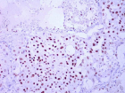
Figure 9
Non-smoking woman with adenocarcinoma of the lung, IV stage with ROS1 mutation and acquired thrombophilia
Immanuels Taivans*, Natalja Senterjakova, Viktors Kozirovskis, Gunta Strazda, Jurijs Nazarovs and Valentina Gordjusina
Published: 04 August, 2021 | Volume 5 - Issue 1 | Pages: 064-072
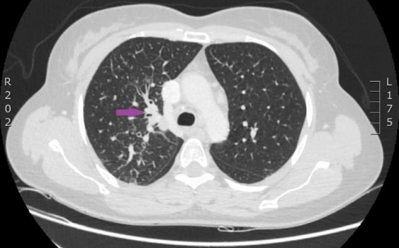
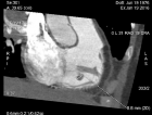
Figure 9:
CT scan of the lungs on 9th of November 2016. Compared with the previous examination, negative changes of the process are seen. Pathological node in the lower lobe of the left lung compared to the previous examination, has increased, its maximal diameter in the axial plane is 24 mm; in S10 paramediastinaly infiltrative focal structure with maximal diameter of 26 mm is present that infiltrates the costal pleura, there is pathological lymph node beside the infiltrative focal structure in the mediastinum, its transverse size in the axial plane is 12 mm. Lymphangitis has progressed in the upper lobe of the right lung; enlarged lymph nodes are present in the mediastinum. Small nodules are present on the right intralobar, costal and diaphragmatic pleura and similar nodules on the diaphragmatic pleura in the left side. (CT scans from Diagnostic Radiology Institute of P.Stradins CUH).
Read Full Article HTML DOI: 10.29328/journal.jprr.1001027 Cite this Article Read Full Article PDF
More Images
Similar Articles
-
Non-smoking woman with adenocarcinoma of the lung, IV stage with ROS1 mutation and acquired thrombophiliaImmanuels Taivans*,Natalja Senterjakova,Viktors Kozirovskis,Gunta Strazda,Jurijs Nazarovs,Valentina Gordjusina. Non-smoking woman with adenocarcinoma of the lung, IV stage with ROS1 mutation and acquired thrombophilia. . 2021 doi: 10.29328/journal.jprr.1001027; 5: 064-072
Recently Viewed
-
Nematophagous Fungus: Pochonia chlamydosporia and Duddingtonia flagrans in the Control of Helminths in Laying Hens (Gallus gallus domesticus) Genus Hy-line Brown - Evaluation and EffectivenessIsabella Allana Ferreira*,Júlia dos Santos Fonseca,Ítalo Stoupa Vieira,Lorendane Millena de Carvalho,Jackson Victor de Araújo. Nematophagous Fungus: Pochonia chlamydosporia and Duddingtonia flagrans in the Control of Helminths in Laying Hens (Gallus gallus domesticus) Genus Hy-line Brown - Evaluation and Effectiveness. Insights Vet Sci. 2025: doi: 10.29328/journal.ivs.1001046; 9: 001-007
-
Biopesticides use on cotton and their harmful effects on human health & environmentPranay Raja Bhad*. Biopesticides use on cotton and their harmful effects on human health & environment. Int J Clin Microbiol Biochem Technol. 2022: doi: 10.29328/journal.ijcmbt.1001025; 5: 005-008
-
Capabilities Approaches Applied to the Homes of the FutureJorge Pablo Aguilar-Zavaleta*. Capabilities Approaches Applied to the Homes of the Future. Ann Civil Environ Eng. 2025: doi: 10.29328/journal.acee.1001081; 9: 066-070
-
The Clinical Pregnancy and Live Birth Following Transfer of One Arrested Embryo: A Case ReportAli Asghar Ghafarizade, Elham Shojafar*, Samira Naderi, Fatemeh Seifi, Alireza Noshad, Zohreh Lavasani, Zahra Kalhori, Elahe Ghadiri. The Clinical Pregnancy and Live Birth Following Transfer of One Arrested Embryo: A Case Report. Clin J Obstet Gynecol. 2024: doi: 10.29328/journal.cjog.1001175; 7: 112-114
-
Intrauterine Therapy with Platelet-Rich Plasma for Persistent Breeding-Induced Endometritis in Mares: A ReviewThiago Magalhães Resende*,Renata Albuquerque de Pino Maranhão,Ana Luisa Soares de Miranda,Lorenzo GTM Segabinazzi,Priscila Fantini. Intrauterine Therapy with Platelet-Rich Plasma for Persistent Breeding-Induced Endometritis in Mares: A Review. Insights Vet Sci. 2024: doi: 10.29328/journal.ivs.1001045; 8: 039-047
Most Viewed
-
Feasibility study of magnetic sensing for detecting single-neuron action potentialsDenis Tonini,Kai Wu,Renata Saha,Jian-Ping Wang*. Feasibility study of magnetic sensing for detecting single-neuron action potentials. Ann Biomed Sci Eng. 2022 doi: 10.29328/journal.abse.1001018; 6: 019-029
-
Evaluation of In vitro and Ex vivo Models for Studying the Effectiveness of Vaginal Drug Systems in Controlling Microbe Infections: A Systematic ReviewMohammad Hossein Karami*, Majid Abdouss*, Mandana Karami. Evaluation of In vitro and Ex vivo Models for Studying the Effectiveness of Vaginal Drug Systems in Controlling Microbe Infections: A Systematic Review. Clin J Obstet Gynecol. 2023 doi: 10.29328/journal.cjog.1001151; 6: 201-215
-
Prospective Coronavirus Liver Effects: Available KnowledgeAvishek Mandal*. Prospective Coronavirus Liver Effects: Available Knowledge. Ann Clin Gastroenterol Hepatol. 2023 doi: 10.29328/journal.acgh.1001039; 7: 001-010
-
Causal Link between Human Blood Metabolites and Asthma: An Investigation Using Mendelian RandomizationYong-Qing Zhu, Xiao-Yan Meng, Jing-Hua Yang*. Causal Link between Human Blood Metabolites and Asthma: An Investigation Using Mendelian Randomization. Arch Asthma Allergy Immunol. 2023 doi: 10.29328/journal.aaai.1001032; 7: 012-022
-
An algorithm to safely manage oral food challenge in an office-based setting for children with multiple food allergiesNathalie Cottel,Aïcha Dieme,Véronique Orcel,Yannick Chantran,Mélisande Bourgoin-Heck,Jocelyne Just. An algorithm to safely manage oral food challenge in an office-based setting for children with multiple food allergies. Arch Asthma Allergy Immunol. 2021 doi: 10.29328/journal.aaai.1001027; 5: 030-037

HSPI: We're glad you're here. Please click "create a new Query" if you are a new visitor to our website and need further information from us.
If you are already a member of our network and need to keep track of any developments regarding a question you have already submitted, click "take me to my Query."


