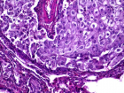
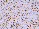
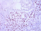
Figure 2
Non-smoking woman with adenocarcinoma of the lung, IV stage with ROS1 mutation and acquired thrombophilia
Immanuels Taivans*, Natalja Senterjakova, Viktors Kozirovskis, Gunta Strazda, Jurijs Nazarovs and Valentina Gordjusina
Published: 04 August, 2021 | Volume 5 - Issue 1 | Pages: 064-072
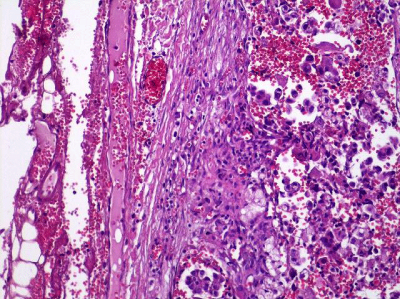
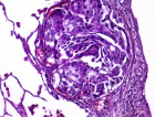
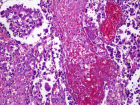
Figure 2:
Poorly differentiated tubular papillary and solid metastatic adenocarcinoma in the lymph node with invasion through the lymph node capsule. Lymphangitis carcinomatosa. (x20, H&E. Dept of Pathology, Medical faculty of the University of Latvia and Institute of Pathology of P. Stradins CUH.
Read Full Article HTML DOI: 10.29328/journal.jprr.1001027 Cite this Article Read Full Article PDF
More Images
Similar Articles
-
Non-smoking woman with adenocarcinoma of the lung, IV stage with ROS1 mutation and acquired thrombophiliaImmanuels Taivans*,Natalja Senterjakova,Viktors Kozirovskis,Gunta Strazda,Jurijs Nazarovs,Valentina Gordjusina. Non-smoking woman with adenocarcinoma of the lung, IV stage with ROS1 mutation and acquired thrombophilia. . 2021 doi: 10.29328/journal.jprr.1001027; 5: 064-072
Recently Viewed
-
Autoantibodies in Autoimmune Addison’s Disease: Why are they Important?Maria Rosaria De Cagna, Norma Notaristefano, Maurizio Schiavone, Gianluca Palatella, Federica Ranù, Carmela Presicci, Valerio Cecinati, Marilina Tampoia*. Autoantibodies in Autoimmune Addison’s Disease: Why are they Important?. Arch Pathol Clin Res. 2024: doi: 10.29328/journal.apcr.1001042; 8: 012-015
-
Acute Gas Toxicity at Work: A Tale of Two Cases with Review of LiteratureRishabh Kumar Singh,Jitender Pratap Singh,Manjari Kishore*,HM Garg. Acute Gas Toxicity at Work: A Tale of Two Cases with Review of Literature. J Forensic Sci Res. 2025: doi: 10.29328/journal.jfsr.1001091; 9: 125-128
-
Cerebral Autoregulation-Directed Therapy in Adults with Non-Traumatic Brain Injury in Neuro-Critical Care: A Scoping ReviewYassine Haimeur*,Mouhssine Doumiri,Mourad Amor. Cerebral Autoregulation-Directed Therapy in Adults with Non-Traumatic Brain Injury in Neuro-Critical Care: A Scoping Review. J Clin Intensive Care Med. 2025: doi: 10.29328/journal.jcicm.1001053; 10: 013-022
-
Experiences of Consumers on the Health Effects of Fake and Adulterated Medicines in NigeriaChijioke M Ofomata, Nkiru N Ezeama, Chinelo Ezejiegu*. Experiences of Consumers on the Health Effects of Fake and Adulterated Medicines in Nigeria. Arch Pharm Pharma Sci. 2024: doi: 10.29328/journal.apps.1001059; 8: 075-081
-
Fetal Bradycardia Caused by Maternal Hypothermia: A Case ReportMuna Alqralleh,Rahma Al-Omari,Shrouq Aldahabi,Doha Abdelbage,Maher Al-Hajjaj*,Lujain Alababesh. Fetal Bradycardia Caused by Maternal Hypothermia: A Case Report. Clin J Obstet Gynecol. 2025: doi: 10.29328/journal.cjog.1001180; 8: 001-002
Most Viewed
-
Feasibility study of magnetic sensing for detecting single-neuron action potentialsDenis Tonini,Kai Wu,Renata Saha,Jian-Ping Wang*. Feasibility study of magnetic sensing for detecting single-neuron action potentials. Ann Biomed Sci Eng. 2022 doi: 10.29328/journal.abse.1001018; 6: 019-029
-
Evaluation of In vitro and Ex vivo Models for Studying the Effectiveness of Vaginal Drug Systems in Controlling Microbe Infections: A Systematic ReviewMohammad Hossein Karami*, Majid Abdouss*, Mandana Karami. Evaluation of In vitro and Ex vivo Models for Studying the Effectiveness of Vaginal Drug Systems in Controlling Microbe Infections: A Systematic Review. Clin J Obstet Gynecol. 2023 doi: 10.29328/journal.cjog.1001151; 6: 201-215
-
Prospective Coronavirus Liver Effects: Available KnowledgeAvishek Mandal*. Prospective Coronavirus Liver Effects: Available Knowledge. Ann Clin Gastroenterol Hepatol. 2023 doi: 10.29328/journal.acgh.1001039; 7: 001-010
-
Causal Link between Human Blood Metabolites and Asthma: An Investigation Using Mendelian RandomizationYong-Qing Zhu, Xiao-Yan Meng, Jing-Hua Yang*. Causal Link between Human Blood Metabolites and Asthma: An Investigation Using Mendelian Randomization. Arch Asthma Allergy Immunol. 2023 doi: 10.29328/journal.aaai.1001032; 7: 012-022
-
An algorithm to safely manage oral food challenge in an office-based setting for children with multiple food allergiesNathalie Cottel,Aïcha Dieme,Véronique Orcel,Yannick Chantran,Mélisande Bourgoin-Heck,Jocelyne Just. An algorithm to safely manage oral food challenge in an office-based setting for children with multiple food allergies. Arch Asthma Allergy Immunol. 2021 doi: 10.29328/journal.aaai.1001027; 5: 030-037

HSPI: We're glad you're here. Please click "create a new Query" if you are a new visitor to our website and need further information from us.
If you are already a member of our network and need to keep track of any developments regarding a question you have already submitted, click "take me to my Query."


