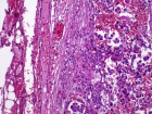
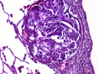
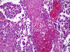
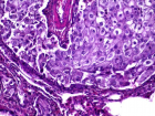
Figure 5
Non-smoking woman with adenocarcinoma of the lung, IV stage with ROS1 mutation and acquired thrombophilia
Immanuels Taivans*, Natalja Senterjakova, Viktors Kozirovskis, Gunta Strazda, Jurijs Nazarovs and Valentina Gordjusina
Published: 04 August, 2021 | Volume 5 - Issue 1 | Pages: 064-072
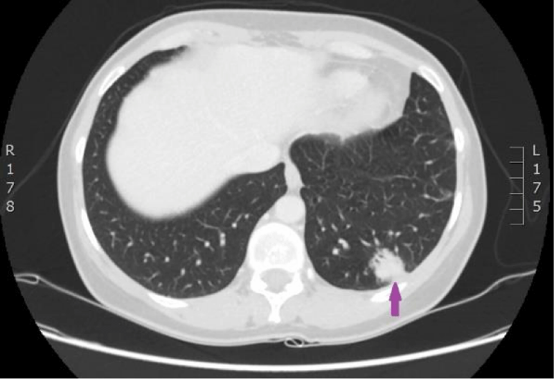
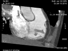
Figure 5:
29.08.2016. Computed tomography of the lungs. Compared with the previous examination, performed on 18.07.2016, the size of the structure in the left lower lobe has increased in all planes (maximal diameter in the axial plane is 23 mm), and new subpleural intrapulmonary lymph nodes (9 and 12 mm) near the mediastinal pleura have appeared. (CT scan from Diagnostic Radiology Institute of P.Stradins CUH).
Read Full Article HTML DOI: 10.29328/journal.jprr.1001027 Cite this Article Read Full Article PDF
More Images
Similar Articles
-
Non-smoking woman with adenocarcinoma of the lung, IV stage with ROS1 mutation and acquired thrombophiliaImmanuels Taivans*,Natalja Senterjakova,Viktors Kozirovskis,Gunta Strazda,Jurijs Nazarovs,Valentina Gordjusina. Non-smoking woman with adenocarcinoma of the lung, IV stage with ROS1 mutation and acquired thrombophilia. . 2021 doi: 10.29328/journal.jprr.1001027; 5: 064-072
Recently Viewed
-
Autoantibodies in Autoimmune Addison’s Disease: Why are they Important?Maria Rosaria De Cagna, Norma Notaristefano, Maurizio Schiavone, Gianluca Palatella, Federica Ranù, Carmela Presicci, Valerio Cecinati, Marilina Tampoia*. Autoantibodies in Autoimmune Addison’s Disease: Why are they Important?. Arch Pathol Clin Res. 2024: doi: 10.29328/journal.apcr.1001042; 8: 012-015
-
Acute Gas Toxicity at Work: A Tale of Two Cases with Review of LiteratureRishabh Kumar Singh,Jitender Pratap Singh,Manjari Kishore*,HM Garg. Acute Gas Toxicity at Work: A Tale of Two Cases with Review of Literature. J Forensic Sci Res. 2025: doi: 10.29328/journal.jfsr.1001091; 9: 125-128
-
Cerebral Autoregulation-Directed Therapy in Adults with Non-Traumatic Brain Injury in Neuro-Critical Care: A Scoping ReviewYassine Haimeur*,Mouhssine Doumiri,Mourad Amor. Cerebral Autoregulation-Directed Therapy in Adults with Non-Traumatic Brain Injury in Neuro-Critical Care: A Scoping Review. J Clin Intensive Care Med. 2025: doi: 10.29328/journal.jcicm.1001053; 10: 013-022
-
Experiences of Consumers on the Health Effects of Fake and Adulterated Medicines in NigeriaChijioke M Ofomata, Nkiru N Ezeama, Chinelo Ezejiegu*. Experiences of Consumers on the Health Effects of Fake and Adulterated Medicines in Nigeria. Arch Pharm Pharma Sci. 2024: doi: 10.29328/journal.apps.1001059; 8: 075-081
-
Fetal Bradycardia Caused by Maternal Hypothermia: A Case ReportMuna Alqralleh,Rahma Al-Omari,Shrouq Aldahabi,Doha Abdelbage,Maher Al-Hajjaj*,Lujain Alababesh. Fetal Bradycardia Caused by Maternal Hypothermia: A Case Report. Clin J Obstet Gynecol. 2025: doi: 10.29328/journal.cjog.1001180; 8: 001-002
Most Viewed
-
Feasibility study of magnetic sensing for detecting single-neuron action potentialsDenis Tonini,Kai Wu,Renata Saha,Jian-Ping Wang*. Feasibility study of magnetic sensing for detecting single-neuron action potentials. Ann Biomed Sci Eng. 2022 doi: 10.29328/journal.abse.1001018; 6: 019-029
-
Evaluation of In vitro and Ex vivo Models for Studying the Effectiveness of Vaginal Drug Systems in Controlling Microbe Infections: A Systematic ReviewMohammad Hossein Karami*, Majid Abdouss*, Mandana Karami. Evaluation of In vitro and Ex vivo Models for Studying the Effectiveness of Vaginal Drug Systems in Controlling Microbe Infections: A Systematic Review. Clin J Obstet Gynecol. 2023 doi: 10.29328/journal.cjog.1001151; 6: 201-215
-
Prospective Coronavirus Liver Effects: Available KnowledgeAvishek Mandal*. Prospective Coronavirus Liver Effects: Available Knowledge. Ann Clin Gastroenterol Hepatol. 2023 doi: 10.29328/journal.acgh.1001039; 7: 001-010
-
Causal Link between Human Blood Metabolites and Asthma: An Investigation Using Mendelian RandomizationYong-Qing Zhu, Xiao-Yan Meng, Jing-Hua Yang*. Causal Link between Human Blood Metabolites and Asthma: An Investigation Using Mendelian Randomization. Arch Asthma Allergy Immunol. 2023 doi: 10.29328/journal.aaai.1001032; 7: 012-022
-
An algorithm to safely manage oral food challenge in an office-based setting for children with multiple food allergiesNathalie Cottel,Aïcha Dieme,Véronique Orcel,Yannick Chantran,Mélisande Bourgoin-Heck,Jocelyne Just. An algorithm to safely manage oral food challenge in an office-based setting for children with multiple food allergies. Arch Asthma Allergy Immunol. 2021 doi: 10.29328/journal.aaai.1001027; 5: 030-037

HSPI: We're glad you're here. Please click "create a new Query" if you are a new visitor to our website and need further information from us.
If you are already a member of our network and need to keep track of any developments regarding a question you have already submitted, click "take me to my Query."


