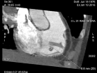
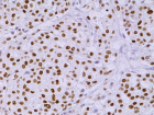
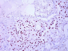
Figure 0
Non-smoking woman with adenocarcinoma of the lung, IV stage with ROS1 mutation and acquired thrombophilia
Immanuels Taivans*, Natalja Senterjakova, Viktors Kozirovskis, Gunta Strazda, Jurijs Nazarovs and Valentina Gordjusina
Published: 04 August, 2021 | Volume 5 - Issue 1 | Pages: 064-072
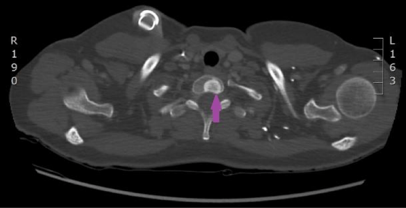
Figure 0:
Computed tomography of the lungs. 18th April 2016. Positive dynamics after therapy: local changes in the basal segment of the left lung have regressed, as well did lymphangitis, and hilar and mediastinal lymphadenopathy. Metastasis to the vertebral body, has been sclerotized after therapy (Illustration from Diagnostic Radiology Institute of P. Stradins CUH).
Read Full Article HTML DOI: 10.29328/journal.jprr.1001027 Cite this Article Read Full Article PDF
More Images
Similar Articles
-
Non-smoking woman with adenocarcinoma of the lung, IV stage with ROS1 mutation and acquired thrombophiliaImmanuels Taivans*,Natalja Senterjakova,Viktors Kozirovskis,Gunta Strazda,Jurijs Nazarovs,Valentina Gordjusina. Non-smoking woman with adenocarcinoma of the lung, IV stage with ROS1 mutation and acquired thrombophilia. . 2021 doi: 10.29328/journal.jprr.1001027; 5: 064-072
Recently Viewed
-
Success Rate and Complications of Endoscopic Deacryocystorhinostomy without Stenting: A Retrospective StudyTulachan B*,Acharya R. Success Rate and Complications of Endoscopic Deacryocystorhinostomy without Stenting: A Retrospective Study. Heighpubs Otolaryngol Rhinol. 2025: doi: 10.29328/journal.hor.1001030; 9: 001-004
-
Assessment of Albino Beech Supremacy to Pigmented Beech Proves to Be A Better Environmental Condition BioindicatorRenata Gagić-Serdar*,Miroslava Marković,Ljubinko Rakonjac,Goran Češljar,Bojan Konatar. Assessment of Albino Beech Supremacy to Pigmented Beech Proves to Be A Better Environmental Condition Bioindicator. Insights Biol Med. 2025: doi: 10.29328/journal.ibm.1001031; 9: 009-015.
-
Rare Locations of Plasma Cell Tumour: A Single-Centre ExperienceVladimir Prandjev,Donika Vezirska,Ivan Kindekov*. Rare Locations of Plasma Cell Tumour: A Single-Centre Experience. J Hematol Clin Res. 2025: doi: 10.29328/journal.jhcr.1001036; 9: 015-019
-
Age Pyramid Assessment of Commercially Important Fishes, Cirrhinus mrigala and Oreochromis niloticus, from the Tropical Yamuna River, IndiaPriyanka Mayank,Amitabh Chandra Dwivedi*. Age Pyramid Assessment of Commercially Important Fishes, Cirrhinus mrigala and Oreochromis niloticus, from the Tropical Yamuna River, India. Insights Biol Med. 2025: doi: 10.29328/journal.ibm.1001029; 9: 001-004
-
Comprehensive Acceptance Testing and Performance Evaluation of the Symbia Intevo Bold SPECT/CT System for Clinical UseSubhash Chand Kheruka*,Naema Al-Maymani,Noura Al-Makhmari,Huda Al-Saidi,Sana Al-Rashdi,Anas Al-Balushi,Anjali Jain,Khulood Al-Riyami,Rashid Al-Sukaiti. Comprehensive Acceptance Testing and Performance Evaluation of the Symbia Intevo Bold SPECT/CT System for Clinical Use. J Radiol Oncol. 2025: doi: 10.29328/journal.jro.1001076; 9: 017-030
Most Viewed
-
Feasibility study of magnetic sensing for detecting single-neuron action potentialsDenis Tonini,Kai Wu,Renata Saha,Jian-Ping Wang*. Feasibility study of magnetic sensing for detecting single-neuron action potentials. Ann Biomed Sci Eng. 2022 doi: 10.29328/journal.abse.1001018; 6: 019-029
-
Evaluation of In vitro and Ex vivo Models for Studying the Effectiveness of Vaginal Drug Systems in Controlling Microbe Infections: A Systematic ReviewMohammad Hossein Karami*, Majid Abdouss*, Mandana Karami. Evaluation of In vitro and Ex vivo Models for Studying the Effectiveness of Vaginal Drug Systems in Controlling Microbe Infections: A Systematic Review. Clin J Obstet Gynecol. 2023 doi: 10.29328/journal.cjog.1001151; 6: 201-215
-
Prospective Coronavirus Liver Effects: Available KnowledgeAvishek Mandal*. Prospective Coronavirus Liver Effects: Available Knowledge. Ann Clin Gastroenterol Hepatol. 2023 doi: 10.29328/journal.acgh.1001039; 7: 001-010
-
Causal Link between Human Blood Metabolites and Asthma: An Investigation Using Mendelian RandomizationYong-Qing Zhu, Xiao-Yan Meng, Jing-Hua Yang*. Causal Link between Human Blood Metabolites and Asthma: An Investigation Using Mendelian Randomization. Arch Asthma Allergy Immunol. 2023 doi: 10.29328/journal.aaai.1001032; 7: 012-022
-
An algorithm to safely manage oral food challenge in an office-based setting for children with multiple food allergiesNathalie Cottel,Aïcha Dieme,Véronique Orcel,Yannick Chantran,Mélisande Bourgoin-Heck,Jocelyne Just. An algorithm to safely manage oral food challenge in an office-based setting for children with multiple food allergies. Arch Asthma Allergy Immunol. 2021 doi: 10.29328/journal.aaai.1001027; 5: 030-037

HSPI: We're glad you're here. Please click "create a new Query" if you are a new visitor to our website and need further information from us.
If you are already a member of our network and need to keep track of any developments regarding a question you have already submitted, click "take me to my Query."


