Figure 8
Non-smoking woman with adenocarcinoma of the lung, IV stage with ROS1 mutation and acquired thrombophilia
Immanuels Taivans*, Natalja Senterjakova, Viktors Kozirovskis, Gunta Strazda, Jurijs Nazarovs and Valentina Gordjusina
Published: 04 August, 2021 | Volume 5 - Issue 1 | Pages: 064-072
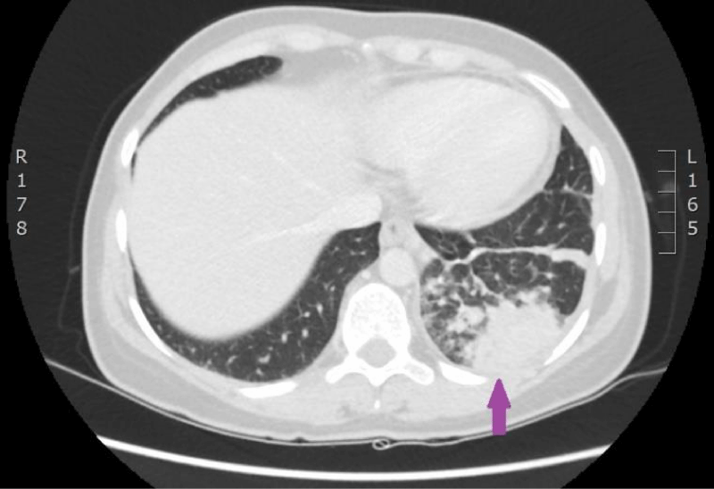
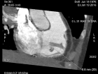
Figure 8:
Computed tomography of the lungs. 4th March 2016. Peripheral adenocarcinoma in the left lower lobe basally, Pathologic infiltrative focus in S9,and S10 segments. Diffuse lymphangitis in the right lung, and in lower lobe of the left lung; bilateral hilar lymphadenopathy of lungs, mediastinum and left cervical area. (Image from Diagnostic Radiology Institute of P.Stradins CUH).
Read Full Article HTML DOI: 10.29328/journal.jprr.1001027 Cite this Article Read Full Article PDF
More Images
Similar Articles
-
Non-smoking woman with adenocarcinoma of the lung, IV stage with ROS1 mutation and acquired thrombophiliaImmanuels Taivans*,Natalja Senterjakova,Viktors Kozirovskis,Gunta Strazda,Jurijs Nazarovs,Valentina Gordjusina. Non-smoking woman with adenocarcinoma of the lung, IV stage with ROS1 mutation and acquired thrombophilia. . 2021 doi: 10.29328/journal.jprr.1001027; 5: 064-072
Recently Viewed
-
Minds after Death: The Expanding Role of Psychological Autopsy in Investigations: A ReviewIshan Jain*,Oindrila Mahapatra,Yogesh Kumar. Minds after Death: The Expanding Role of Psychological Autopsy in Investigations: A Review. J Forensic Sci Res. 2025: doi: 10.29328/journal.jfsr.1001096; 9: 155-0
-
Comparison of Body Fat Percentage and BMI in Pre-hypertensive and Hypertensive Female College Students of West TripuraPuja Saha,Satyapriya Roy,Susmita Banik,Sonali Das,Shilpi Saha*. Comparison of Body Fat Percentage and BMI in Pre-hypertensive and Hypertensive Female College Students of West Tripura. J Adv Pediatr Child Health. 2025: doi: 10.29328/journal.japch.1001070; 8: 001-006
-
Harmonizing Artificial Intelligence Governance; A Model for Regulating a High-risk Categories and Applications in Clinical Pathology: The Evidence and some ConcernsMaxwell Omabe*. Harmonizing Artificial Intelligence Governance; A Model for Regulating a High-risk Categories and Applications in Clinical Pathology: The Evidence and some Concerns. Arch Pathol Clin Res. 2024: doi: 10.29328/journal.apcr.1001040; 8: 001-005
-
Parallelism of the Evolution of Social Insects and Humans: A HypothesisAV Makrushin*. Parallelism of the Evolution of Social Insects and Humans: A Hypothesis. Arch Psychiatr Ment Health. 2024: doi: 10.29328/journal.apmh.1001055; 8: 038-040
-
The Power of Inner Dialogue: The Impact of Self-Talk Techniques on Athlete PerformanceVeysel Temel*. The Power of Inner Dialogue: The Impact of Self-Talk Techniques on Athlete Performance. Arch Clin Exp Orthop. 2025: doi: 10.29328/journal.aceo.1001021; 9: 001-003
Most Viewed
-
Feasibility study of magnetic sensing for detecting single-neuron action potentialsDenis Tonini,Kai Wu,Renata Saha,Jian-Ping Wang*. Feasibility study of magnetic sensing for detecting single-neuron action potentials. Ann Biomed Sci Eng. 2022 doi: 10.29328/journal.abse.1001018; 6: 019-029
-
Evaluation of In vitro and Ex vivo Models for Studying the Effectiveness of Vaginal Drug Systems in Controlling Microbe Infections: A Systematic ReviewMohammad Hossein Karami*, Majid Abdouss*, Mandana Karami. Evaluation of In vitro and Ex vivo Models for Studying the Effectiveness of Vaginal Drug Systems in Controlling Microbe Infections: A Systematic Review. Clin J Obstet Gynecol. 2023 doi: 10.29328/journal.cjog.1001151; 6: 201-215
-
Prospective Coronavirus Liver Effects: Available KnowledgeAvishek Mandal*. Prospective Coronavirus Liver Effects: Available Knowledge. Ann Clin Gastroenterol Hepatol. 2023 doi: 10.29328/journal.acgh.1001039; 7: 001-010
-
Causal Link between Human Blood Metabolites and Asthma: An Investigation Using Mendelian RandomizationYong-Qing Zhu, Xiao-Yan Meng, Jing-Hua Yang*. Causal Link between Human Blood Metabolites and Asthma: An Investigation Using Mendelian Randomization. Arch Asthma Allergy Immunol. 2023 doi: 10.29328/journal.aaai.1001032; 7: 012-022
-
An algorithm to safely manage oral food challenge in an office-based setting for children with multiple food allergiesNathalie Cottel,Aïcha Dieme,Véronique Orcel,Yannick Chantran,Mélisande Bourgoin-Heck,Jocelyne Just. An algorithm to safely manage oral food challenge in an office-based setting for children with multiple food allergies. Arch Asthma Allergy Immunol. 2021 doi: 10.29328/journal.aaai.1001027; 5: 030-037

HSPI: We're glad you're here. Please click "create a new Query" if you are a new visitor to our website and need further information from us.
If you are already a member of our network and need to keep track of any developments regarding a question you have already submitted, click "take me to my Query."


