
More Information
Submitted: October 07, 2026 | Accepted: October 14, 2026 | Published: October 16, 2026
Citation: Chen LR. Breathing Movement of Asthmatic Airways. J Pulmonol Respir Res. 2026; 10(2): 26-42. Available from:
https://dx.doi.org/10.29328/journal.jprr.1000180
DOI: 10.29328/journal.jprr.1000180
Copyright license: © 2026 Chen LR. This is an open access article distributed under the Creative Commons Attribution License, which permits unrestricted use, distribution, and reproduction in any medium, provided the original work is properly cited.
Keywords: Controlled breathing (steady-slow breathing); Controlled coughing; Obstructed points; Targeted respiratory maneuvers (active breathing therapy); Conventional procedure; Targeted procedure
Abbreviations: CB: Controlled Breathing; CC: Controlled Coughing; OPs: Obstructed Points; Fully OPs: Meaning Points that are totally blocked; Partial OPs: OPs were opened during inspiration and closed during expiration

Breathing Movement of Asthmatic Airways
Li Ren Chen*
Internal Medicine, Guangdan Worker Hospital, Guangzhou, Guangdong, China
*Corresponding author: Li Ren Chen, Internal Medicine, Guangdan Worker Hospital, Guangzhou, Guangdong, China, Email: [email protected]
Airway narrowing is the primary pathophysiological abnormality in asthma. Mucus plugs within the lumen remain a critical and prevalent cause of airway obstruction and contain inflammatory secretions that must be removed and drained. Therefore, the clearance of mucus plugs is vital for effective treatment and disrupts the vicious cycle of chronic airway remodeling in my clinical practice. Mucus retention and subsequent obstruction occur both in patients with reduced or restricted ventilation and those experiencing respiratory arrest. To investigate the mechanical forces driving mucus transport, I conducted experiments using custom-built tube-balloon models to observe the dynamics of air movement and mucus displacement within the tubes and balloons during various cycles of breathing and coughing. Clinical application of these experimental findings suggests: their efficacy in asthma management; airway respiratory movement is the primary force driving secretion transport; targeted respiratory maneuvers facilitate the clearance of trapped air and mucus plugs, particularly within the distal small airways. This approach allows for the gradual reduction and discontinuation of bronchodilators while preventing acute relapses and chronic exacerbations. Furthermore, a subtle wheezing detected during gentle, prolonged expiration between attacks and immediately following the subsidence of the acute episode is identified as a key clinical indicator of distal mucus blockage. In addition, suppression of the pharyngeal cough reflex promotes continuous cephalad displacement of secretions. Finally, the use of acupuncture or acupoint injection as an adjunct therapy significantly enhances the therapeutic effects of the breathing exercises.
The basic pathophysiologic abnormality that determines the functional and symptomatic status of an asthmatic patient is airway narrowing [1].
In the management of asthma, in addition to the recognized factors of edema, inflammatory cell infiltration, congestion, bronchospasms, and remodeling of the airway wall, mucus plugging within the lumen remains a critical and prevalent cause of airway obstruction [1-3]. Bronchographic studies demonstrated “that airway plugging is widespread between attacks and is prominent in patients with fatal asthma [2]”. These obstructions are present “in airways of all sizes distal to the second order bronchi, but the most striking changes are observed in airways around 5 mm in diameter [2]”. This specific level of the bronchial tree is particularly vulnerable, as it represents the site where proximal airway collapse peaks at the end of normal expiration typically in healthy individuals [2]. Similarly, recent reports have indicated that the percentage of obstructions caused by mucus plugs is notably greater in fourth- and fifth- generation bronchi (which represent airways centered around 5 mm in diameter) than in other generations of bronchi [4]. Numerous investigators and current reviews continue to emphasize the pivotal role of small airways mucus plugging in the pathophysiology of asthma [5,6].
It is important to identify the primary forces driving the transport and removal of these secretions. Current theory suggests that airway secretions are removed through three biological mechanisms: absorption, lymphatic drainage, and phagocytosis. Any residue that is not absorbed is transported cephalad (upward) toward the epiglottis, where it enters the esophagus and gastrointestinal tract. This transport is facilitated by ciliary activity, smooth muscle contraction, postural drainage, and the respiratory movement of the airway walls [7].
In clinical practice, the retention of airway secretions—leading to significant obstruction—is most prevalent in three patient populations: (1) those with diminished respiratory excursion, such as bedridden patients without primary lung disease; (2) those with restricted thoracic expansion, which is common in cases of chest trauma; and (3) those with absent spontaneous respiration (e.g., respiratory failure). In the latter case, the cessation of airway movement halts natural mucus transport, necessitating mechanical ventilation and manual suctioning for clearance [8]. Logically, these observations suggest that ciliary activity and smooth muscle contraction alone are insufficient for complete secretion transport when breathing movement is restricted. Conversely, enhancing the dynamic movement of the airway wall—such as through encouraged deep breathing and coughing—effectively prevents secretion accumulation and clears trapped mucus [8]. Therefore, it is proposed that ciliary action primarily serves to decouple (separate) secretions from the airway wall and act as a one-way valve through upward beating. This action facilitates mucus transport, but the primary mechanical impetus remains the respiratory movement of the airway walls.
In the study: the displacement of mucus and plugs in a non-ciliated environment is investigated by constructing a physical respiratory model (tube-balloon models) consisting of synthetic tubing (representing the tracheobronchial tree), balloons (representing the alveoli), and a manually simulated chest cavity, which is for purposes to confirm: 1). respiratory movement of the airway walls is the primary mechanical impetus for transportation and removal of airway mucus; and 2) the clearance of mucus and plugs from obstructed airways is essential for effective asthma management. The rationale is:
As I was a medical student observing a patient with an acute asthma attack, I noted that while the primary symptoms subsided after treatment, the patient continuously expectorated a large amount of mucoid sputum—a feature my instructor emphasized as classic. In contrast, chronic asthma patients typically exhibited long-term prolonged expiration accompanied by distinct phlegm sounds. These clinical observations led to my foundational hypothesis: mucous plugging was an inherent characteristic of asthma. Later, bronchography has shown that “airway plugging is widespread between asthmatic attacks as well as being prominent in patients dying of asthma [2].”
And, exactly, the airway is infiltrated with inflammatory cells and asthmatic mucus, and plugs inherently consist of inflammatory secretions. Therefore, my rationale in 1971 drew a historical parallel to the 18th-century surgical doctrine for treating abscesses: “incision, pus evacuation, and drainage” —a single decisive stroke that brought surgeons renown, provided that unobstructed drainage was strictly maintained. Plugging mucus with inflammatory secretions in the airway must be treated with the same surgical rigor.
Custom-built tube-balloon models (Patent-United States application number: 19/751,083 filed on 07/22/2026)
I. Structural design: The general architecture of the models is designed to replicate the human tracheobronchial tree and alveoli as perceived by macroscopic (naked eye) observation.
II. Experimental observation: The models facilitate repeated observations of the cephalad displacement of the dyed mucus and plugs, airflow dynamics, and mechanical movement of the model walls. These dynamics are evaluated in both healthy and mucus-plugged asthmatic simulations during various cycles of breathing and coughing.
III. The plugged tubes are designed to simulate asthmatic airways after bronchodilator treatment (excluding instances of paradoxical responses or other risks associated with β2-adrenoceptor agonists [9]): in the asthmatic airway,
1. Failure of biological clearance: The mechanisms of absorption, lymphatic drainage, and phagocytosis are compromised because of significant vascular leakage [10].
2. Ciliary denudation: Ciliary functions are neutralized as cilia are frequently shed in the asthmatic airway [14]; and
3. Reduced smooth muscle efficacy: Airway smooth muscle exhibits diminished force generation because of the following:
(1) Mechanical overdistension: Airway dilation resulting from the accumulation of excessive air, mucus, and plugs;
(2) Increased afterload: The heightened mechanical load imposed by dense, intraluminal mucus plugging; and
(3) Structural alterations: smooth muscle hypertrophy and hyperplasia.
This collective mechanical failure further exacerbates the difficulty of airway clearance, resulting in respiratory wall movement being the remaining primary force for transport.
Experimental observations focus on different simulated breathing and coughing processes: characterizing the biomechanics and functional movement of healthy tube-balloon walls and specific mechanical features and dysfunctions in mucus-plugged asthmatic models. By extrapolating these findings, this study sought to infer the physiological features and function of healthy airway wall movement and the mechanical failures and structural dysfunctions that occur in obstructed airways. The ultimate goal of this research is to develop methods that facilitate the transport and clearance of mucus and plugs from obstructed airways, thereby restoring effective ventilation to the affected pulmonary segments.
Evaluation of custom-built tube-balloon models
In these experimental trials, dynamic wall movements consist specifically of: longitudinal elongation (rhythmic axial stretching, creating differential displacement of mucus that facilitated displacement from distal, smaller caliber tubes into proximal, larger caliber tubes), radial expansion (triggering tension that facilitated separated action of mucus, and spread of mucus-valve action), Radial contraction (thickened action of mucus generating a “valve action”, and enhanced “squeeze-pump effect”), and axial shortening (piston-like displacement generating a significant “squeeze-pump effect”). These targeted respiratory maneuvers within the plugged-model experiments utilize trapped or redistributed air as a pneumatic medium. This generates positive intraluminal pressure of a large volume of air and high-velocity airflow, facilitating intraluminal expansion, dislodgment, and expulsion of the mucus plugs (at the beginning of CC). These maneuvers also leverage intrapleural pressure fluctuations to create pressure gradients, radial traction, and suction forces, which activate air redistribution and drive the transport and clearance of mucus, particularly from the bronchiolar tubes. Proper maneuvers, maintaining steady, slow breathing after the CC of conventional procedures or targeted procedures, are essential for establishing optimal baseline conditions; this stability facilitates the removal of trapped air and secretions through subsequent CC of conventional procedures or targeted procedures [15,16]. Such breathing patterns represent the functional reserve capacity of the model’s respiratory movements, which are critical for effective clearance. These findings suggest that mechanical deformation of the airway wall is a primary physical driver for mucus clearance, potentially redefining the functional hierarchy between cilia and mechanical respiratory movement.
Experimental observations were evaluated and applied to clinical practices:
Clinical application of these experimental observations confirms the efficacy of specific targeted respiratory maneuvers. These basic breathing practices include: controlled breathing (CB: steady-slow respiration characteristic of resting states); controlled coughing (CC: brief, low-impact expulsive efforts); sustained deep inspiration (a steady-slow inhalation followed by a 3–5 second pause to maximize thoracic expansion); and sustained expiratory effort (after deep inspiration, a 3–5 second isometric contraction of the intercostal and abdominal muscles against a closed glottis).
These maneuvers achieve therapeutic effects through gradual cephalad displacement and clearance of mucus-containing inflammatory secretions from the airways, particularly from small airways. This clearance facilitates the alleviation and full reversal of airway narrowing, allows for gradual tapering and eventual discontinuation of bronchodilator therapy, and prevents acute relapses and chronic exacerbations. Furthermore, patients benefit from acupuncture and acupoint injection, which appears to modulate airway smooth muscle tone via nervous system stimulation. This adjunct therapy facilitates the recruitment of secondary respiratory muscle groups, thereby improving the precision and proficiency of the breathing exercises.
The two treatment methods, each of which requires prolonged periods and collaboration with the other, shorten the treatment course to prevent acute relapses of asthma and the exacerbation of chronic asthma, which is worthy of further study and application. It is especially evident that the localization and clearance of mucus and plugs in the small airways required more in-depth investigation because I depend only on experiments involving models (subtle wheezing was detected during gentle, prolonged exhalation), (confirmed by) previous experience in clinical practice, and analyses of anatomy.
Evidence from physical modeling, clinical practice, and physiological theory suggests that airway wall movement is the fundamental driver of respiratory health. The coordinated action of respiratory movement of the tracheobronchial tree and mechanical deformation of the alveoli, integrated with ciliary activity and smooth muscle tone, may be collectively defined as tracheobronchial and alveolar motility—a vital functional component of overall airway motility.
Mechanical deformation of the airway wall serves as the primary physical driver of mucus clearance [8]. Targeted respiratory maneuvers utilize the vast functional reserve capacity of respiratory movement of the airway wall and have proven effective in clinical practice for dislodging and clearing obstructions [15,16]. Ultimately, the mobilization and clearance of plugging mucus-containing inflammatory secretion—from the airway, particularly from the small airways—are critical interventions for achieving a comprehensive therapeutic effect in asthma management.
Experiments on custom-built tube-balloon models 1971-1973
Figure 1 Respiratory system in the experiments involving custom-built tube-balloon models.
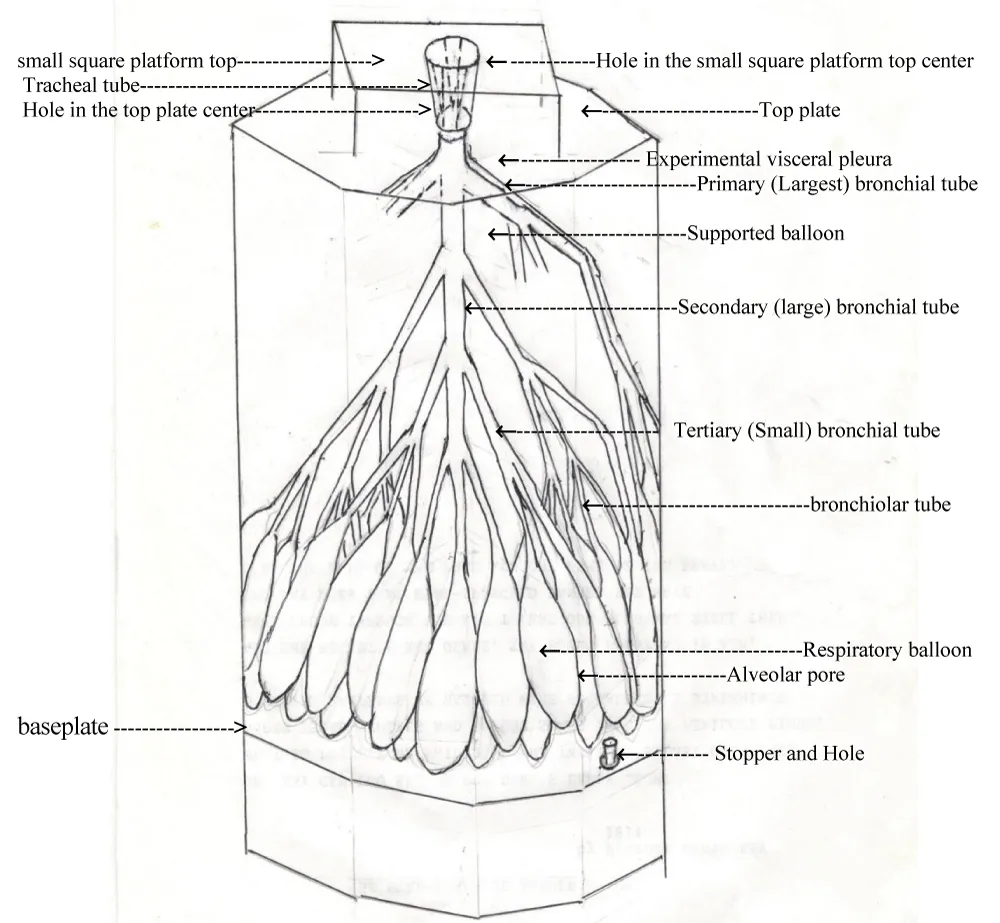
Figure 1: Respiratory system in the experiments involving custom-built tube-balloon models.
Tube-Balloon Apparatus: The experimental apparatus comprises four custom-built tube-balloon models of approximately equal dimensions. The primary components—colorless, transparent, flexible polymer tubing and balloons—were sourced from repurposed landfill materials. These models are housed within a simulated thoracic cavity (experimental thorax).
Mechanical Operation: Respiratory cycles are simulated using a mobile baseplate acting as an experimental diaphragm. During inspiration, the baseplate is retracted (pulled downward) to create negative pressure; during expiration, it is advanced (pushed upward). In the healthy (unobstructed) model, the exhaled volume is approximately 600 mL during simulated normal expiration and approximately 1200 mL during deep expiratory maneuvers.
The experimental setup consisted of four custom-built tube-balloon systems, each comprising an experimental lung (tracheobronchial tubes and balloons) housed within a simulated thorax (see Figure: Respiratory System in Experiments of Custom-built Tube-balloon Models). Synthetic mucus and plugs were instilled into the models to visualize the dynamics of airflow, secretion transport, and the mechanical response of the tube-balloon structures. Observations were conducted across various phases of respiration and coughing to evaluate mucus transport in unobstructed models and the clearance of obstructions in asthmatic simulations. The ultimate objective was to determine how these mechanical interventions could be applied in clinical practice to alleviate and fully reverse airway narrowing in patients with asthma.
Experimental model
The experimental lung (comprising the tracheal tube assembly) consisted of a large outer balloon, representing the visceral pleura, which encapsulated a smaller, pre-inflated internal balloon. This internal balloon was ligated at the neck to maintain a fixed air volume following the removal of its rim.
The interstitial space between these structures contained a network of bronchial tubes and respiratory balloons (alveolar analogs), all of which are submerged in a specific volume of paraffin oil. The bronchial tubes branched in a root-like morphological pattern and were distributed across the surface of the internal balloon, with the exception of its base. The respiratory balloons were positioned specifically beneath the inferior aspect (bottom) of the internal balloon.
Finally, the neck of the large outer balloon was hermetically secured to the distal end of the tracheal tube using a high-strength adhesive.
Tubes (airways)
The diameters of the synthetic tubes were tapered progressively from the proximal segment of the tracheal tube to the distal terminations of the bronchiolar tubes. Similarly, the wall thickness of the model components steadily decreased from the proximal segment of the tracheal wall to the distal tips of the respiratory balloons [17].
Tracheal and bronchial dimensions:
Tracheal Tube: Measuring 12 cm in length, the tracheal tube with a proximal inner diameter of 2 cm extended vertically into the experimental lung.
Primary (Largest) Bronchial Tubes: The distal end of the trachea branched into three primary tubes, each 6 cm in length.
Secondary (Large) Bronchial Tubes: Each primary tube connected to three secondary tubes, which were also 6 cm in length.
Tertiary (small) Bronchial Tubes: Each secondary tube branched into three tertiary tubes, each measuring 10 cm in length.
Bronchiolar Tubes: Finally, each tertiary tube connected to three bronchiolar tubes, each measuring 8 cm in length.
The experimental tubes were categorized into two distinct segments: the non-collapsed ‘Large-Tubes’ and the collapsed ‘Small-Tubes’ at the end of normal expiration in unobstructed models.
Large-Tubes (Large-Airways)
The “Large-Tubes” contained cervical tracheal tube located between the elevated platform and the top plate gasket and intrapleural Large-Tubes located between the top plate gasket and the experimental lung were defined as the non-collapsed tubes remaining at the end of normal expiration. This category encompassed the tracheal tube, primary and secondary bronchial tubes, and the larger-caliber small bronchial tubes, which extended from the proximal end of the tracheal tube to a specific (functional) level of the bronchial tree. This distal boundary corresponded to the midpoint of the small bronchial tubes—the specific point where distal tubes (Small-Tubes) collapse terminated at the conclusion of normal expiration.
The mechanical behavior of these segments differed significantly on the basis of their anatomical location: Intrapleural Large-Tubes exhibited dynamic changes synchronized with the respiratory cycle, elongating and dilating during inspiration, while recoiling, compressing, and shortening during expiration. In contrast, the cervical portion of the trachea (represented in the model as the segment above the thoracic superior boundary) demonstrated restricted movement, undergoing only axial elongation during inspiration and shortening during expiration.
Small-Tubes (Small-Airways)
The Small-Tubes consisted of the smaller caliber tertiary bronchial tubes (5 cm in length) and bronchiolar tubes. In this model, the bronchiolar tubes are represented by the necks of the respiratory balloons (simulating the alveoli). This section spanned from the distal boundary of the large tubes to the terminal end of the bronchiolar tubes, which maintained a minimum internal diameter of 2 mm to ensure experimental patency and connection stability. These segments exhibited dynamic changes synchronized with the respiratory cycle, elongating, opening, and dilating during inspiration, but recoiling, compressing, collapsing, and shortening during expiration.
Respiratory balloons (alveoli)
Abbreviation: balloon (respiratory balloon)
The experimental lung was constructed using 81 droplet-shaped (respiratory) balloons, with the balloon necks serving as the bronchiolar tubes. Each balloon had a total volume of 11 mL and was preloaded with 3 mL of gelatinized starch suspension to represent simulated airway secretions. Adjacent balloons were connected at 3 to 4 small interfaces (1 cm in diameter) using adhesive. Within these contact points, 3 to 4 micro-perforations (less than 1 mm in diameter) were established to simulate the Pores of Kohn. Mechanically, these structures were observed to recoil, compress, wrinkle, and collapse during expiration but elongate, open, and dilate during inspiration.
For the experimental thorax, see Figure small square platform
The experimental thorax was a hexagonal prism composed of six colorless and transparent glass panels, a hexagonal wooden top plate, and a hexagonal wooden baseplate (simulating the diaphragm). A small square platform (6 cm in height) was mounted at the center of the top plate.
At the center of the top plate, an aperture was fitted with a soft gasket to serve as a low-friction bearing. The outer surface of the middle tracheal tube and the inner surface of this gasket were lubricated with paraffin oil. The experimental lung assembly was then inserted, with the proximal end of the tracheal tube passing upward through the gasket and securely bonded to a central hole in the elevated platform.
Respiratory simulation
The baseplate functioned as a simulated diaphragm. To calibrate the system, a stopper in the baseplate is removed to set the initial volume and then reinserted to ensure an airtight seal. Respiration is simulated by pulling the baseplate downward (inspiration) and pushing it upward (expiration). In the normal models, the exhaled air volume is approximately 600 mL during tidal expiration and 1200 mL during deep expiration.
Establishment of reference marks: Reference markers were established on the thoracic wall to indicate the baseplate levels corresponding to the following physiological states:
End-tidal inspiration: The position of the baseplate when the balloons were filled with a standard volume of air.
EnSteady-slow inspiration: The position of the baseplate when the balloons were filled with air volume that was less than that of end-tidal inspiration.
Steady-slow deep inspiration: The position reached when the balloon volume exceeded tidal levels. This was measured during three distinct maneuvers: steady-slow deep inspiration, sustained steady-slow deep inspiration, and simulated labored asthmatic inspiration.
End-tidal expiration: The level at which the small tubes collapsed to their specific collapse point within the bronchial tree.
End steady-slow expiration: The level at which the small tubes collapsed but did not reach their specific collapse point within the bronchial tree.
End steady-slow deep expiration: The level at which airway collapse progressed proximally to the midpoint of the secondary bronchial tube. This was recorded during steady-slow deep expiration, sustained steady-slow deep expiration, and simulated labored asthmatic expiration.
Special materials and instrumentation
Simulated Secretions (Mucus): Two concentrations were prepared: (a) standard mucus (~1% starch concentration) and (b) high-viscosity mucus (~2–3% starch concentration). These samples were prepared by mixing glutinous rice flour with distilled water until smooth, and then heating to a boil to achieve gelatinization.
Mucus plugs: Plugs were fabricated by extruding a 5% starch batter into boiling water using various syringes and needle gauges. The resulting plugs ranged from 2–10 mm in diameter and 5 mm–5 cm in length. Plugs were harvested once they reached buoyancy at the boiling point. Contrast Agents (Dyes): 2% mercurochrome (red) (from 1971 to 1973) and 2% crystal violet (purple) solutions are utilized for visual tracking of flow dynamics within the models.
Glass funnel: A standard laboratory funnel consisting of a conical body and an integrated stem. Alveolar Cannula: A flexible plastic tube used for the targeted injection of simulated mucus into individual respiratory balloons.
Multidiameter cannulae: Various flexible tubes of differing diameters used to precisely deposit mucus and plugs at specific levels within the bronchial tree.
Endotracheal collection system: A straight plastic tube (< 2 cm diameter, 10 cm length) connected to a 2000 mL collapsed plastic reservoir. This system was used to collect total expiratory air volume during various simulated breathing events in both normal and asthmatic models.
Volumetric measurement: A 100 mL syringe was used to precisely quantify the collected expiratory air volumes.
Normal models
In normal models, simulated mucus stored within the respiratory balloons was transported upward, coating the inner luminal surfaces of the tubes before being expelled from the upper cervical tracheal segment during tidal respiration.
Experimental procedures
To load the model, the stem of a glass funnel was inserted into the tracheal lumen. Simulated mucus was poured into the funnel and drawn into the distal tubes and balloons by slowly displacing the baseplate downward (simulated inspiration).
Following the filling phase, respiratory maneuvers commenced. Initial inspiratory airflow and gravity caused any excess mucus to migrate distally into the balloons. During subsequent tidal respiration (normal breathing), a thin mucous film was transported proximally toward the upper cervical trachea only. This mechanism of clearance during tidal breathing proved superior to that of other simulated respiratory patterns.
Observation of clearance
To facilitate the observation of mucus expulsion, the proximal end of the tracheal tube was detached from the central tabletop aperture. The proximal 4 cm of the tube was incised longitudinally into four uniform strips, which were then secured to the perimeter of the tabletop opening (4 cm in diameter). This modification allowed clear visualization of mucus being transported upward and out of the upper cervical tracheal segment during the respiratory cycle.
Mucus-plugged asthmatic models
(Mild to moderate) Mucus-Plugged asthmatic models
Definition of airway passages
A bronchial passage was defined as a continuous conduit extending from an individual respiratory balloon to the tracheal tube. In this model, these passages were categorized into four states of patency:
- Unaffected passages: Containing only a thin film of standard mucus.
- Non-Obstructed Plugged Passages : Containing a thin mucous film and small plugs that did not compromise the airway lumen.
- Partially obstructed passages: Containing high-viscosity mucus and larger plugs that resulted in partial luminal obstruction during inspiration, and complete luminal obstruction during expiration.
- Completely obstructed passages: Containing high-viscosity mucus and larger plugs that resulted in complete luminal obstruction.
Experimental procedures:
1. To establish approximately 18 to 20 unaffected bronchial passages, standard mucus was injected into selected bronchial tubes and their corresponding balloons using an alveolar cannula.
2. To create approximately 18 to 20 non-obstructed bronchial passages, standard mucus was injected, followed by the placement of “smaller plugs” at various anatomical levels. These plugs were sized to ensure that they did not create OPs (Obstructed Points) or significant resistance.
3. To simulate approximately 40 partially obstructed bronchial passages, high-viscosity mucus was injected into these remaining bronchial units. Specialized cannulae were subsequently used to deposit mucus and plugs of increasing diameters into segments ranging from the distal bronchioles to the proximal large bronchi. The plug diameters were carefully selected to ensure only partial obstruction of the tube lumen. This assembly constituted the mild-to-moderate mucus-plugged asthmatic model.
Status asthmaticus (severe mucus-plugged) models
Experimental procedures:
To simulate severe asthma exacerbation, the mild-to-moderate model was modified through a specialized mechanical protocol. Initially, all unaffected passages, non-obstructed plugged passages, and a small subset of partially obstructed passages were physically clipped (clamped). Subsequently, labored asthmatic breathing was simulated. Under these high-pressure conditions, the remaining partially obstructed passages progressed to complete luminal occlusion (total obstruction). Upon releasing the initially clipped tubes, the model entered a state of severe, acute exacerbation, representing Asthmaticus Status.
Observation of alveolar heterogeneity:
Throughout the experiments, the Status Asthmaticus models consistently demonstrated significant heterogeneity in balloon (alveolar) volume. In each trial, the 81-balloon array exhibited the following distribution:
- Severe hyperinflation: approximately 30 excessively inflated balloons.
- Partial Inflation (>50% volume): 8 to 10 balloons.
- Minimal Inflation (<50% volume): 4 to 5 balloons.
- Atelectasis (Collapse): 4 to 6 balloons containing no air.
Models with isolated bronchiolar mucus plugging definition
This model was designed to simulate obstruction localized strictly to the distal airways. It featured high-viscosity mucus within selected balloons and a combination of high-viscosity mucus and plugs within their corresponding bronchiolar tubes.
Experimental procedures:
To establish localized plugging, an alveolar cannula was used to inject high-viscosity mucus into a subset of balloons. A fine-gauge cannula was subsequently used to deposit viscous mucus and a graded series of plugs (ranging from smallest to largest diameter) into the corresponding bronchiolar tubes, spanning from the distal to the proximal bronchiolar junctions. All other tubes and balloons are injected with standard mucus to maintain baseline patency.
Observation of occlusion
Following the initiation of any simulated respiratory pattern, all the plugs in the targeted bronchiolar tubes resulted in complete luminal occlusion. This protocol successfully established a model of isolated bronchiolar obstruction.
Clinical practices (daily outpatient and respiratory training work)
Targeted respiratory maneuvers (active breathing therapy) of models (and airways)
Conventional procedures:
1) Controlled breathing (CB): Steady-slow breathing
It is a maneuver that mimics resting respiration. The respiratory cycle was characterized by regular, smooth movements with a rate, velocity, and depth intentionally maintained below those of standard tidal respiration. In the experimental simulations, the expiratory volume during CB was approximately 400 mL. This was significantly greater (by approximately 50 mL) than the expiratory volume recorded during simulated labored asthma breathing (~350 mL), suggesting improved ventilation efficiency.
2) Controlled coughing (CC)
Controlled coughing was defined as a low-intensity, short cough initiated following deep inspiration. The maneuver required the immediate cessation of expiratory effort as soon as the initial mild, short cough occurred and was followed by CB.
Targeted procedures
This therapeutic protocol consisted of a three-stage mechanical sequence designed to optimize airway clearance:
Stage 1: Deep inspiration (maximizing airway caliber)
To maximize the internal diameter of the airways and open the largest full OPs and the collapsed airways and alveoli, the patient performed one of two maneuvers:
1) Steady-slow deep inspiration: Inhaled air volume exceeded the tidal volume, or
2) Sustained deep inspiration (inspiration hold): Following steady-slow deep inhalation, the expansion of the thoracic cavity was maintained for 3 to 5 seconds to ensure maximum bronchial and alveolar recruitment.
Stage 2: Sustained expiratory effort (pressure augmentation)
Maintaining upward pressure on the baseplate and the proximal end of the tracheal tube was occluded (a mild, sustained expiratory muscle contraction against a closed glottis) for 3–5 seconds.
Stage 3: Controlled cough (kinetic clearance)
Following maintaining upward pressure on the baseplate (the sustained expiratory effort against a closed glottis and once mentally and physically ready) and opening of the proximal end of the tracheal tube (the glottis was opened) for an explosive release of air in the form of a mild, short cough.
Auxiliary procedures
Pharyngeal cough stimulation (usually this stimulation persists for only seconds)
Clinical observation: I identified a novel therapeutic approach: the active inhibition of pharyngeal cough stimulation. Suppressing the immediate cough reflex appeared to promote the continuous cephalad (proximal) displacement of mucus and plugs, although the precise underlying mechanism remains unclear.wing distribution:
Management of pharyngeal stimulation:
When patients experienced a localized “urge to cough” in the pharynx, the following suppression methods were employed:
Method 1: Behavioral inhibition and relaxation
The patient was instructed to remain calm and achieve total body relaxation. They must tolerate the stimulation without immediate reaction, utilizing CB or targeted procedures (as previously described). It was critical to strictly avoid premature or spontaneous coughing, particularly a paroxysmal bout of consecutive coughs initiated without any preceding or intervening inspiration.
Method 2: Chemical suppression (Iodine)
Patients applied two drops of 5% iodine solution (or 2% iodine tincture) to each side of the middle third of the tongue. The solution was retained or gargled in the pharyngeal area to provide local stimulation/suppression without being swallowed.
Method 3: Topical anesthesia (Lidocaine)
Patients applied two drops of 1% lidocaine solution to each side of the middle third of the tongue. Like Method 2, the solution was gargled or retained in the pharynx to achieve rapid desensitization.
Clinical response and mucus clearance:
Latency: Under Method 1 or 2, pharyngeal stimulation typically subsides within seconds. Method 3 results in the near-immediate disappearance of the cough urge.
Clearance: Following the successful suppression of the cough reflex, mucus plugs were observed to migrate to the large bronchi or lower trachea within minutes. Once the plugs reach these proximal segments, CC is utilized for final expectoration.
Acupuncture treatment (traditional Chinese medicine)
Prescription: Acupuncture or acupoint injection, targeting 1 to 2 points of the lung channel of Hand-Taiyin per session, was administered 1 to 2 times per week for several months as needed.
Acupuncture:
Method: Filiform needles were used to puncture specific points along the Lung Channel of Hand-Taiyin. For excess-type syndromes (characterized by acute, prominent symptoms), the reducing method was applied. For deficiency-type syndromes (which are characterized by physical weakness and chronic fatigue), the reinforcing method was applied [19].
Precautions: Strict attention must be given to the proper depth and direction of needle insertion to ensure safety and efficacy.
Acupoint injection:
Method: A volume of 0.5-1.0 mL of 0.9% sodium chloride or 1% lidocaine was injected into one or two points along the Lung Channel of Hand-Taiyin. Treatments were administered once per week for several months as clinically indicated. The therapeutic effects observed were comparable to those of traditional acupuncture.
Precautions: Strict adherence to the proper depth and direction of needle insertion was essential.
Therapeutic management and manipulation
Clinical practices include integrated targeted respiratory maneuvers, auxiliary procedures (as needed), and acupuncture or acupoint injection.
Interictal Periods (Between Asthma Attacks)
To prevent acute relapses of asthma, the following protocol was implemented:
Targeted respiratory maneuvers: These were performed consistently. Patients performed targeted procedures at least twice every 20 minutes during waking hours. Alternatively, patients performed targeted procedures twice whenever they remembered, and twice upon awakening.
Auxiliary procedures: These procedures were performed as needed.
Acupuncture or acupoint injection: Treatment was administered 1 to 2 times per week for several months as needed.
Acute attacks
Regardless of whether patients received conventional medical treatment, the following protocol was strictly followed:
Breathing control: Patients maintained steady, slow breathing to mitigate the distress associated with labored asthmatic breathing.
Airway clearance: Targeted respiratory maneuvers were performed consistently, with a focus on targeted procedures and auxiliary procedures as needed to clear mucus and plugs from the airways.
Fatigue management: If exhaustion occurred after repeated targeted procedures, patients switched to CB for a recovery period.
Chronic asthma
The primary therapeutic goal was the effective clearance of mucus plugs from the airways. This was achieved through the consistent application of targeted respiratory maneuvers, specifically targeted procedures and special auxiliary procedures as needed. Adherence to these protocols led to progressive symptom relief and the gradual prevention of asthma exacerbations.
In a specific case of chronic asthma, asthma symptoms were completely resolved after 24 months of persistent targeted respiratory maneuvers, particularly through the rigorous application of targeted procedures (patients performed targeted procedures at least 2 times every 20 minutes); mucus and plugs are not necessarily expectorated; following the completion of the targeted procedure’s closed cycle, mucus and plugs were mobilized and displaced cephalad to the larger bronchi or lower trachea within minutes, and ultimately expectorated. These maneuvers were essential protocols that required long-term maintenance to remove mucus and plugs to prevent the exacerbation of chronic asthma and ultimately resolve asthma symptoms.
Experiments using 6 normal models
During normal breathing, a thin mucous film was observed transporting cephalad (upward) along the inner surface of the model walls, eventually exiting through the upper cervical tracheal tube. Compared with all other breathing techniques, this method demonstrates superior transport efficiency. Across all trials, the respiratory rate, velocity, intensity, and depth, as well as the mechanical movements of the model walls, the speed and volume of mucus transport, and the total exhaled air volume remained consistent.
26 Mucus-plugged asthma model experiments
This study utilized 26 mucus-plugged asthmatic models, which were categorized into 12 mild-to-moderate models and 14 status asthmaticus (severe) models.
The results demonstrated that even the largest fully occluded OPs, involving collapsed tubes and balloons, were successfully reopened. Through the application of targeted respiratory maneuvers, mucus and plugs were mobilized from the smaller-caliber tubes into the larger-caliber tubes without inducing secondary occlusions. Ultimately, total clearance of all mucus and plugs was achieved across all 26 experimental models.
Six models with isolated bronchiolar mucus plugging
In each experiment, the following results were observed: subtle wheezing was detected in the plugged bronchiolar tubes during gentle, prolonged exhalation. By utilizing targeted procedures exclusively, mucus and plugs were mobilized and transported cephalad into the larger-caliber tubes without inducing secondary occlusions, slowly and gradually. These mucus and plugs were subsequently cleared from these larger tubes using targeted respiratory maneuvers.
Clinical practices
Three treatment methods were applied to 3 types of clinical manifestations of 54 asthmatic patients. See Table 1.
| Table 1: Summary of Clinical practices. (The number enclosed in parentheses is the case number.) | |||||
| Treated methods | Results | ||||
| Diseases | 1. between asthmatic attacks | 2. Uncontrolled acute asthmatic attacks following bronchodilator | 3. Chronic asthma | Total cases of followed up for 5 years | |
| Patients’ age | 30 to 50 | ||||
| Method 1. targeted respiratory maneuvers, & auxiliary procedures as need | cases | (4) | (4) | (4) | 12 |
| Expelled mucus & plugs | Yes (1), and not (3). | Yes (3), and not (1). | Yes (4), | ||
| discontinued bronchodilator use | N/A | Not 4. | (4) decreased dose | ||
| prevention | Yes (8), ceased after 6 months. | Yes (4) prevented exacerbation after 6 months | |||
| Method 2. Treatment of acupuncture or acupoint injection |
3 | 3 | 3 | 9 | |
| Expelled mucus & plugs | Not (3), | Yes (1), and Not (2). | Yes (3) | ||
| discontinued bronchodilator use | N/A | Not (3), | (3) decreased dose. | ||
| prevention | Yes, (6) Relapses decreased after 6 months. |
(3) prevented exacerbation after 6 months. | |||
| Method 3. Combination therapy: targeted respiratory maneuvers, auxiliary procedures as need, & treatment of acupuncture or acupoint injection | Cases | (4) + method 1. (4) + method 2. (3) | (4) + method 1. (4) + method 2. (3) | (4) + method 1. (4) + method 2. (3) | 33 |
| Expelled mucus & plugs | Yes (5) and not (6). | Yes (11). Fully reverse airway narrowing | Yes (11). | ||
| discontinued bronchodilator use | N/A | N/A | Decreased dose (11) | ||
| prevention | Yes (22), ceased within 3 months. | (10) exacerbation was prevented after 3 months and (1) symptoms ceased after24 months. | |||
| Conventional treatment | Cases | (10) | (10) | (10) | 30 |
| Patients’ conditions | Being controlled (6) and relapsed (4). | Being controlled (8) and exacerbate (2). | Being controlled (8) and exacerbated (2). | ||
Table: Method 3 is the best choice for the prevention of acute asthma relapse and exacerbation of chronic asthma. Mucus plugging is an important cause of airway narrowing in patients with asthma. Targeted respiratory maneuvers are important measures for achieving a therapeutic effect gradually: mucus and plugs (particularly in the small airways) are transported proximally to the larger airways to alleviate and fully reverse airway narrowing, gradually reduce and eventually discontinue bronchodilator use, and prevent acute relapses of asthma and exacerbations of chronic asthma. Treatment with acupuncture or acupoint injection is also therapeutically effective in patients with asthma. To achieve therapeutic effects, the two treatments need prolonged periods separately, and a combined regimen of acupuncture or acupoint injection helps improve the therapeutic effect of targeted respiratory maneuvers and shorten the course of treatment: 1) acute asthma relapses are ceased within 3 months gradually in patients without subtle wheezing during gentle, prolonged exhalation between attacks and immediately following the subsidence of acute asthma episodes, and 2) exacerbations of chronic asthma are prevented after 3 months. In chronic asthma, only one patient achieved the best result because this was the only patient who administered strictly targeted respiratory maneuvers, particularly targeted procedures, and auxiliary procedures as needed, and acupuncture or acupoint injection, which was potentially promising for chronic asthma patients.
Results of experiments on custom-built tube-balloon models
Observations in 6 normal model experiments
Transport efficiency: At a respiratory rate of 16 to 18 breaths per minute, normal tidal breathing movements transported mucus cephalad (upward), exiting through the upper cervical tracheal tube. This method proved superior to other breathing patterns, achieving a transport velocity of 6 to 8 mm/min.
Coughing: Effective mucus expulsion (both upward and external) was successful achieved through common coughing.
Definition of failure: Uncontrolled coughing—defined as continuous coughing for several seconds without prior inspiration—failed to achieve effective clearance.
Experiments on 26 mucus-plugged asthmatic models:
Phase I: Initial clearance: Initially, by utilizing conventional procedures, some of the largest severely occluded OPs and collapsed tubes and balloons were reopened. Mucus and plugs from these reopened or partially occluded tubes, and from tubes without occlusions, were gradually transported cephalad into the larger-caliber tubes without inducing secondary occlusions. These mucus and plugs were then cleared via CC of conventional procedures.
Phase II: Targeted intervention: By subsequently utilizing targeted procedures exclusively, the remaining largest fully OPs and collapsed tubes and balloons, which had been refractory (resistant) to conventional procedures, began to open slowly; and the residual mucus and plugs in the tubes, particularly within the bronchiolar tubes, were mobilized and transported cephalad into the larger caliber tubes slowly and gradually.
Phase III: Final clearance: To ensure safety, this mobilization process was conducted gradually to avoid secondary occlusions. Ultimately, all remaining mucus and plugs were successfully removed from these larger-caliber tubes using CC of conventional procedures or targeted procedures as needed.
The transport speed and volume of the mucus and plugs, as well as the expiratory air volume, varied according to the severity of the obstruction and the air volume within the smaller-caliber tubes and/or the corresponding balloons behind the OPs. While the baseline expiratory air volume was derived from the unaffected and unobstructed bronchial passages, the incremental increase in expiratory air volume was directly linked to the clearance of mucus and plugs. Furthermore, the increase in the ventilated air volume was contingent upon the increasing number of unaffected and unobstructed bronchial passages.
In 6 models with isolated bronchiolar mucus plugging
Mucous plugging was exacerbated, leading to full obstruction in the bronchiolar tubes under various breathing patterns, particularly labored asthmatic breathing. Subtle wheezing was detected during gentle, prolonged exhalation. By utilizing targeted procedures exclusively, the mucus and plugs were mobilized and transported cephalad into the larger-caliber tubes (albeit with difficulty) without causing secondary occlusions, slowly and gradually. These mucus and plugs were subsequently cleared from the larger-caliber tubes using targeted respiratory maneuvers.
Results of clinical practices (see Table)
Method 1: This approach effectively cleared mucus and plugs in Diseases 2. & 3. In Disease 3, bronchodilator dosages were gradually reduced; however, achieving an optimal therapeutic effect required at least six months of treatment.
Method 2: This method proved insufficient during the early stage of treatment. Mucus and plugs were transported upward during Diseases 2. & 3. The therapeutic benefits—specifically, the minimization of bronchodilator dose in Disease 3—became apparent after 6 months.
Method 3: Acupuncture or acupoint injection significantly enhanced the patients’ ability to perform targeted respiratory maneuvers and auxiliary procedures. This approach accelerated the course of treatment, leading to a progressively superior therapeutic effect. In all 22 cases involving Disease 1 (11 cases) & Disease 2 (11 cases), acute relapse ceased within three months. Additionally, exacerbations of chronic asthma were successfully prevented after the three-month mark in Disease 3 (11cases).
Notably, one patient with chronic asthma achieved an exceptional therapeutic outcome because of his strict adherence to a consistent regimen of targeted respiratory maneuvers, specifically targeted procedures and auxiliary procedures focused on airway clearance. The targeted procedures were typically performed at least 2 times every 20 minutes. The patient reported: “When my symptoms exacerbated, I did not take a bronchodilator or increase bronchodilator dose and instead performed targeted procedures intensely (the targeted procedures were typically performed 2 times every 10 minutes at least). My symptoms were relieved after even a small amount of mucus was expectorated.” This case may provide significant hope for other chronic asthma patients.
Mucus plugging was a primary cause of airway narrowing in patients with asthma. Consequently, targeted respiratory maneuvers served as an essential measure for achieving gradual therapeutic effects.
These targeted respiratory maneuvers facilitated the cephalad transport of mucus and plugs in the airways, particularly within the small airways, and led to a reduction in and eventual discontinuation of bronchodilator use to alleviate and fully reverse airway narrowing gradually. Furthermore, auxiliary procedures promoted the continuous displacement of the mucus and plugs. Eventually, acute relapse and exacerbation of chronic asthma were prevented over a six-month period.
Patients with asthma experienced therapeutic improvements in airway narrowing when treated with acupuncture or acupoint injection. Both treatments required prolonged periods when administered separately, and the combined regimen enhanced the therapeutic effect and significantly shortened the treatment course. This combined approach gradually prevented the following: 1) acute relapses within 2 to 3 months in the patients: no subtle wheezing was detected during gentle, prolonged expiration between attacks and immediately following the subsidence of the acute episode; and 2) exacerbation of chronic asthma after 3 months. In the treatment of chronic asthma, one patient achieved complete cessation of asthma symptoms after 24 months.
Experiments involving custom-built tube-balloon models
Breathing movement of custom-built tube-balloon models and mucus transport.
The features and functions of movements of the normal model walls during normal breathing.
Inspiration
At the onset of inspiration, the lung model underwent sequential elongation (elongated action). The balloons elongate first, followed by the small-caliber tubes and finally, the large-caliber tubes. This sequence generates pressure from the folding walls, forcing the cephalad displacement of mucus within the collapsed Small-Tubes; however, the mucus in the balloons remains stationary, as the pressure from the elongating balloon walls is insufficient to drive cephalad movement. Simultaneously (at the onset of inspiration), dilation of the distal segments of the Large-Tubes occurs alongside the opening and dilation of the proximal Small-Tubes, and a specific volume of expelled mucus (retained mucus left from the end of the previous expiration) in the distal Large-Tubes flows downward and spreads onto the inner luminal surfaces of the newly opening and dilating proximal Small-Tubes.
Subsequently, the opening and dilation occur from the larger Small-Tubes to the balloons, and the mucus spreads onto the inner surfaces of the opening and dilating Small-Tubes and balloons (acting as a valve). After the collapsed Small-Tubes and balloons are opened and dilated completely, the intrapleural Large-Tubes undergo rapid and intensive elongation and dilation: 1) the mucus on the inner surface including the residual expelled-mucus in the distal portion of the Large-Tubes is sheared and separated from the wall, this separation results from the change in the inner superficial area of the (elongating and dilating) tube wall; and 2) triggering an effect of differential displacement of the mucus in the intrapleural Large-Tubes because the smaller-caliber tubes are elongated downward earlier than the larger-caliber tubes, and the elongated length of the smaller-caliber tubes is greater than that of the larger-caliber tubes.
Inspiration concludes with the elongation of the cervical tracheal tube, which increased its original length by approximately 2 cm. Some residual expelled mucus (retained mucus) remains in the distal portion of the Large-Tubes, as the rapid and intensive elongation and dilation of the intrapleural large tubes fail to propel the mucus cephalad.
Expiration
At the onset of expiration, air flows out of the balloons because the resistance of the balloon walls to intrapleural pressure was lowest in the model group. As the internal air volume gradually decreases, the walls of the intrapleural tubes and balloons are recoiled and compressed. This process forces the thin mucus film on the inner surface of the lumen of the tubes and balloons to accumulate into a thickened layer—thickened action of mucus (a “valve action”). Subsequently, mucus on the inner surface of the balloon lumen detaches and falls because of wrinkling of the balloon walls.
As the balloon air is depleted, the intrapleural pressure transmitted through the lung (parenchyma) becomes sufficient to overcome both the declining transpulmonary pressure and the mechanical rigidity of the walls.7 Consequently, sequential collapse (the “expelled action”) initiates at the balloons (which prevented the upward secretion of the balloon contents) and extends to the proximal Small-Tubes, driven by the varying wall resistances to the intrapleural pressure. In this mechanism, the lower collapsed segment functions as a “pushing piston”, whereas the upper non-collapsed segment acts as a “piston ring”. This coordinated action effectively expels a specific volume of mucus (expelled motion of mucus) into the distal portions of the Large-Tubes.
Following the collapse of the proximal Small-Tubes, the intrapleural Large-Tubes undergo rapid and intensive compression and shortening, along with the shortening of collapsed Small-Tubes and the increasing wrinkling of balloons. Consequently, in the intrapleural Large-Tubes: the smaller-caliber tubes are compressed and shortened earlier than the larger-caliber tubes; particularly, the shortened length of the smaller-caliber tubes is greater than that of the larger-caliber tubes. The smaller-caliber tubes function as a “pushing piston”, whereas the larger-caliber tubes serve as a “piston ring”, and the exhaled airflow forms the “central core” of the tubes, collectively contributing to a “piston-like effect”. This rapid, intensive shortening and compression of the Large-Tubes provide the optimal driving force for mucus clearance (expulsive action).
Expiration concludes with shortening of the cervical tracheal tube, reducing its original length by approximately 0.5 cm. Any residual expelled mucus in the distal segments of the Large-Tubes, which is not propelled cephalad by rapid, intensive compression and shortening of the intrapleural Large-Tubes, remains as retained mucus in the smallest-caliber sections of the Large-Tubes at the end of expiration.
During breathing, mucus transport is limited to the upper cervical tracheal tube. This occurs because the respiratory movement of the cervical tracheal tube is restricted to elongation and shortening, with these length changes being more pronounced in the smaller-caliber cervical tracheal tube than in the larger ones [20]. whereas the intrapleural tubes and balloons also undergo a cycle of opening, dilation, recoil, compression, wrinkling, and collapse; consequently, the longitudinal changes in the tube length work in tandem with diameter fluctuations to facilitate effective mucus transport in the tubes.
Targeted respiratory maneuvers of the model (active breathing therapy)
These proper breathing exercises were designed to prolong positive physiological effects, avoid premature narrowing, closure, or collapse of the corresponding larger-caliber tubes proximal to OPs, and ensure the transport of trapped air, mucus, and plugs.
Conventional procedures: CB-Controlled Breathing (also known as Steady-slow respiration)
Steady-slow inspiration: This technique prevents or reduces the gravitational (downward) migration of mucus and plugs. It facilitates the reopening of some of the largest fully OPs through radial expansion, while also re-expanding some collapsed tubes and balloons through tractive force. Consequently, inhaled air passes through these newly opened or partially opened OPs into smaller-caliber tubes and/or their corresponding balloons. This increases the air volume in the small-caliber tubes distal to the OPs, thereby enhancing the efficacy of subsequent conventional and targeted procedures.
Steady-slow expiration: It reopens some of the largest fully OPs and re-expands some collapsed tubes and balloons, dislodges plugged mucus, and facilitates their upward mobilization. By avoiding excessive expiratory effort, it delays the premature narrowing, closure, or collapse of the larger caliber tubes proximal to OPs, and mitigates dynamic tube compression that occurs even beyond the initial phase of expiration.
CC (controlled coughing): It consists of brief, low-impact (mild, short) coughing following inspiration and once mentally and physically ready. The objective of this “short-pulse” technique is to expectorate mucus and plugs from only the tubes of the larger bronchi and lower trachea. And expiratory effort must stop immediately as soon as the initial mild short cough occurs and be followed by CB, which prevents premature narrowing, closure, or collapse of the larger tubes proximal to the OPs and inhalation of mobilized mucus, and maintains the optimal conditions around the OPs.
Targeted procedures
Sustained or only a steady-slow deep inspiration first: Sustained steady-slow deep inspiration is more effective than a simple steady-slow deep inspiration. In this technique, tractive action is exerted on the model walls because of thoracic expansion. This tractive force acts upon the outer walls of both (1) fully OPs, facilitating their reopening; and (2) collapsed tubes and balloons, the resulting dilation of the lumen in these previously collapsed structures generating intracavitary negative pressure (suction action); this pressure gradient draws air from two primary sources: 1) higher-caliber or smaller-caliber tubes via their respective OPs, and 2) adjacent balloons via alveolar pores (Pores of Kohn), facilitating their re-expansion. Consequently, the likelihood of human atelectasis developing may be decreased [8].
Sustained expiratory effort: No reduction was observed in tube diameter or balloon volume. This was attributed to air-filled lumens and insufficient expiratory pressure to deflate the compliant tubes and balloons. This sustained expiratory effort triggers a redistribution of air that reopens the fully OPs and re-expands collapsed tubes and balloons. Specifically, air from inflated smaller-caliber tubes is forced through the larger OPs into larger-caliber tubes that are the smaller-caliber tubes distal to the (larger or) largest fully OPs, which is a critical prerequisite for enhancing the efficacy of subsequent CB or CC. Furthermore, air is driven from 1) inflated tubes through their relative OPs into the collapsed tubes, and 2) inflated balloons through the alveolar pores (Pores of Kohn) into adjacent underinflated or collapsed balloons. This collateral ventilation re-expands the collapsed tubes and balloons and may decrease the likelihood of human atelectasis.
Finally, once mentally and physically ready, perform
CC (of target procedures): This method is superior in clearance efficiency to CC of the conventional procedure. At the beginning of CC, an explosive release of air 1) mechanically dilates the tube lumen and converts the fully OPs into opened or partial OPs through radial expansion, 2) expectorates mucus and plugs in the tubes of the larger bronchi and lower trachea to the exterior, 3) generates sufficient kinetic energy to dislodge deep-seated mucus cephalad, particularly from the bronchiolar tubes, and 4) forces air into the small-caliber tubes distal to OPs. At the very least, it initiates progressive expansion and dislodgement, enabling the systematic clearance of mucus and plugs. Following the CC of targeted procedures, the patients should transition to steady, slow breathing. This prevents premature narrowing, closure, or collapse of the larger tubes proximal to the OPs and the accidental deep inhalation of mobilized mucus, ensuring that the plugging mucus does not recede into the distal tubes and maintaining the optimal conditions around the OPs before subsequent conventional or targeted procedures. Furthermore, these targeted procedures help suppress intense pharyngeal irritation, reducing the urge for unproductive, reflexive coughing.
Mucus plugging in bronchiolar tubes typically leads to complete obstruction during any breathing except for CC and CB, which severely impairs transport. Only targeted procedures effectively increase the bronchiolar lumen diameter and the air volume in the small caliber bronchiolar tubes and/or corresponding balloons distal to OPs, and enhance the capacity of the following CC and CB. Consequently, it progressively facilitates the clearance of mucus in the bronchiolar tubules, albeit with difficulty. This approach is significantly more effective than CC of conventional procedures.
Biomechanical mechanism of tube-balloon clearance
The superior efficacy of the targeted procedures observed in experimental models is attributed to distinct fluid dynamic mechanisms. During sustained deep inspiration, the expansion of the thorax maximizes the transmural pressure (Ptm), creating a radial distending force that exceeds the critical opening pressure of fully occluded OPs. This is followed by sustained expiratory effort, increasing the air volume within the smaller-caliber tubes and/or their corresponding balloons distal to the OPs. This process is facilitated by collateral ventilation (as mentioned above), which promotes the reopening of OPs and the re-expansion of collapsed tubes and balloons. Consequently, CC of targeted procedures overcomes the viscoelastic adhesion of mucus. This leads to the effective cephalad displacement of mucus plugs, particularly from bronchiolar tubes. These biomechanical interactions explain how, compared with CC of conventional procedures, targeted procedures successfully modulate the internal physical environment of the tube-balloon to achieve superior clearance.
Dysfunction of respiratory movement of mucus-plugged asthmatic models
In the asthma model, smaller-caliber tubes that were already partially obstructed by mucus plugs became completely obstructed at the beginning of expiration. This led to the premature narrowing, closure, or collapse of the corresponding larger-caliber tubes. This phenomenon was particularly evident during asthmatic expiration and occurring even beyond the initial phase of the normal expiratory cycle [14]. These dynamics characterize the wall movement of mucus-plugged tubes, which results in mechanical dysfunction of the tube walls during expiration: Mucus and plugs fail to be transported upward in the corresponding tubes even during normal expiration; instead, they accumulate at the OPs and the plugging tubes. During labored asthma breathing, inhaled air passes OPs to enter their smaller-caliber tubes and/or corresponding balloons during inspiration, but remains trapped during exhalation. These create a ‘one-way valve’ mechanism, resulting in the excessive accumulation of air (hyperinflation), mucus, and plugs within the obstructed tubes and balloons.
Mechanisms of mucus retention and gravitational migration in models
On the basis of experimental models: gravitational (downward) migration of mucus during inspiration occurred within the model lumen, particularly around specific levels of the bronchial tree and tube bifurcations; mucus retention was observed in the distal segments of the large tubes at the end of inspiration and expiration. These result in a localized plugging tendency, driven by the structural design of the tube-balloon models, ventilation patterns, and the properties of the experimental materials—phenomena that appear to mirror the dynamics in the human airways [11].
Model optimization and refinement: balancing anatomical fidelity and experimental stability
In the early stages of model development, researchers attempted to use finer tubing (distal diameter of 1 mm) to achieve higher anatomical fidelity. However, this approach faced significant technical hurdles, including unstable bonding at tube junctions, difficulties in embolus placement, and the disproportionately large volume of the experimental lung. Ultimately, the pilot model failed to demonstrate the intended experimental outcomes, as it could not effectively showcase how luminal narrowing could be alleviated and fully reversed through appropriate interventions. This failure was primarily attributed to the complexities of establishing varied operating pressures and managing OPs (obstructed points) occluding passages. Consequently, the model was refined to a distal diameter threshold of 2 mm. This adjustment is a strategic compromise to ensure experimental reproducibility and mechanical stability. Future studies utilizing advanced microfluidic materials may allow for even smaller-scale simulations to further refine these observations. Consequently, the distal diameter of the 1 mm tubing was deemed impractical for simulating dynamic airway responses.
Clinical practices see Table
The clinical observations discussed herein were derived from routine outpatient respiratory consultations and standard breathing management during daily medical practice. All non-pharmacological interventions—specifically the targeted respiratory maneuvers and breath control techniques—were implemented purely as safe, standard, and non-invasive physiological training for patient care. No experimental drugs, novel invasive procedures, or standard-care-modifying trial protocols were involved. To strictly safeguard patient privacy, all relevant clinical data and observational records have been completely anonymized, with all direct and indirect personal identifiers removed in accordance with standard medical ethics guidelines.
Several limitations of this study must be acknowledged, primarily stemming from its historical and real-world origin dating back several decades in remote mountainous and resource-limited health settings. Due to the lack of local laboratory and diagnostic infrastructure (such as spirometry, peak flow meters, and imaging) at the time, as well as socioeconomic constraints affecting patient compliance with complex testing, objective pre- and post-treatment physiological measurements were not available; long-term adherence to routine systemic medications or complex testing was frequently unfeasible. Evaluation of efficacy was inherently pragmatic; the assessment of therapeutic efficacy relied necessarily on pragmatic, real-world clinical endpoints—specifically, significant reduction or complete cessation of acute attacks and exacerbations of chronic asthma and functional recovery observed in daily life and work environments, which were directly observable by patients, their families, and colleagues in their work environments. Therefore, the findings presented here should be interpreted as preliminary, historical observations from real-world clinical practice rather than controlled trial data, highlighting the need for future validation using modern objective assessments.
In my clinical practice (daily outpatient work), outcomes of asthma management were reported on the basis of clinical improvement or the complete resolution of symptoms, and the reduction or cessation of bronchodilator use. Most patients experienced therapeutic relief following the clearance of airway mucus and plugs. Owing to socioeconomic factors and personal preferences, many patients declined laboratory tests, pulmonary function testing, X-rays, and skin testing. Furthermore, many were unable to adhere to daily systemic medications or long-term medical advice because of their family circumstances. Consequently, I lacked the necessary clinical data to perform a formal analysis of patients’ underlying pathogenesis. To make more accurate observations and reports, I went through a difficult process.
These patients only used bronchodilators during acute attacks or when symptoms worsened, and they typically sought my help only when those medications failed to provide relief. Under these circumstances, I focused on patient education and recommended self-managed active breathing therapy, alongside treatments such as acupuncture or acupoint injections.
Research, involving both custom-built tube-balloon models and clinical practice, had focused exclusively on the clearance of mucus and plugs from obstructed tubes and airways. While this study was specific in scope, it addressed a critical issue: airway obstruction. The clearance of plugging mucus contained inflammatory secretions, which effectively removes a significant contributor to the disease and may help resolve airway inflammation, thereby enhancing the overall therapeutic effect in patients with asthma?
Expectoration of mucus and plug in method 3 was observed in only five patients in disease 1, and in method 1, only one patient in disease 1. It is possible that other patients failed to notice when minute amounts of mucus were transported from airways, particularly peripheral small airways, to the epiglottis.
3 treatment procedures in clinical practice for asthma:
1. Targeted respiratory maneuvers and auxiliary procedures as needed (active breathing therapy)
Targeted respiratory maneuvers:
Controlled respiration: Patients with asthma must maintain steady, slow breathing to mitigate the distress of labored asthma breathing, exacerbated by further airway narrowing, delay and reduce premature narrowing, closure, or collapse of the larger airways proximal to OPs, facilitate plugging mucus cephalad displacement, increase expiratory air volume, and extend the duration of the positive physiological effects of CB and CC.
CC
The expectoration of mucus and plugs is dependent on the CC of conventional procedures or targeted procedures. The therapeutic impact on asthma is characterized by the significant removal of plugging mucus containing inflammatory secretions. CC is the best drainage of inflammation.
Proper use of CC in conventional procedures or targeted procedures: Common coughing often produces audible phlegm sounds, yet the mucus remains difficult to transport upward. In such cases, continued application of CCs in targeted procedures may initially yield no sound or expectoration. However, by maintaining the CB for several minutes, the phlegm was gradually mobilized into the larger caliber airways, such as the major bronchi or lower trachea. If expectoration did not occur after this period, a second series of targeted procedures would typically facilitate the successful clearance of the phlegm. And it was not necessary to take vigorously short coughs (that was more effective to dislodge deep-seated mucus) because of the patient’s physical strength and tolerance. The CC of conventional procedures or targeted procedures is performed only when the patient is mentally and physically ready.
Executing untimely and inappropriate coughing: (as opposed to CC) in response to pharyngeal stimulation often results in labored but ineffective expectoration and exacerbation of airway obstruction. While a portion of the mucus and plugs is expelled, the remaining secretions fail to migrate toward the larger bronchi or lower trachea once the pharyngeal stimuli subside.
Auxiliary procedures
Inhibition of pharyngeal coughing stimulation: Coughing is often triggered by the displacement of mucus and plugs, which stimulates nerve receptors within the bronchial mucosa and, in turn, elicits a reflex stimulation in the pharynx. Untimely or inappropriate coughing prevents mucous cephalad displacement and leads to mucus plugging. Suppression of this pharyngeal cough reflex promotes the continuous cephalad displacement (upward movement) of mucus and plugs. By inhibiting this reflex, once the pharyngeal stimulation has subsided, mucus and plugs can be transported to the larger-caliber airways—including the primary bronchi and lower trachea—within minutes; they are ultimately expelled via the CC of conventional procedure. The underlying physiological mechanisms for this suppression effect remain to be fully elucidated.
However, in cases of persistent and uncontrollable coughing stimulation, the continued application of targeted procedures facilitates the steady, cephalad propulsion of the mucus; this gradual upward movement of the mucus and plugs can, in turn, mitigate local irritation and help suppress the cough reflex.
2. Treatment of acupuncture or acupoint injection
This regulates the nervous system to modulate airway smooth muscle tone. This mechanism facilitates the recruitment of appropriate muscle groups, thereby improving the precision and proficiency of proper breathing exercises.
3. Targeted therapy
Acupuncture or acupoint injection serves as an effective adjunct to targeted respiratory maneuvers. This combination potentiates the therapeutic effects in asthma patients, although the underlying physiological mechanisms remain to be fully elucidated.
An important finding involved the mechanism of asthmatic wheezing
In the models with isolated bronchiolar mucus plugging: Typically, mucus plugs in the bronchiolar tubes cause complete obstruction during any respiration except for CC and CB, particularly during labored asthmatic breathing. During normal expiration, most of the airflow bypasses the obstructed segments, exiting via unaffected tubes and non-obstructed plugged tubes. At the end of normal expiration, because the Small-Tubes and balloons have already collapsed, the air and air pressure remain within the smaller bronchiolar tubes and/or corresponding balloons distal to the OPs. As further expiratory effort and the intrapleural pressure is transmitted through the simulated lung and acts on the corresponding distal bronchiolar tubes and balloons to force a trace volume of air through the OPs that emits a subtle wheezing. Interestingly, this subtle wheezing often disappears during labored expiration, as the high expiratory pressure transmitted through the simulated lung likely compresses the external walls of the OPs, further sealing the bronchiolar obstruction.
To determine whether mucus plugs remained in the small airways, in experimental observations of custom-built tube-balloon models, subtle wheezing was detected during gentle, prolonged expiration. This finding was validated in clinical practice, where patients either report the sensation or wheezing is detected via auscultation during gentle, prolonged expiration between asthmatic attacks and immediately following the subsidence of the acute episode. Through anatomic and physiologic analysis, consequently, this specific wheezing is identified as a key diagnostic marker for mucus blockage within the distal airways.
In a normal airway, during prolonged deep expiration, the posterior membranous portion collapses far into the lumen of the trachea and large-caliber bronchi, narrowing the lumen into thin slits that also experience whistling [14-21], and in the asthma model, premature narrowing, closure, or collapse of the larger-caliber tubes proximal to the corresponding mucus plugging of smaller-caliber tubes occurs at the beginning of expiration; this phenomenon occurs even beyond the initial phase of the normal expiratory cycle and is particularly evident during asthmatic expiration, which narrows the lumen of the trachea and large-caliber bronchi into thin slits and also experiences whistling. The gravitational (downward) migration of mucus and plugs happens even during normal inspiration. Consequently, in asthmatic airways, breathing should be steady, slow breathing instead of asthmatic breathing, and the cough must be a short cough.
In general, the clearance of plugging mucus containing inflammatory secretions from obstructed airways through targeted respiratory maneuvers is a recognized method for treating asthma.
Implications and suggestions
Mucus plugging is an inherent characteristic of asthma, prevailing both between acute attacks and prominently in fatal cases. Airway mucus plugging remains a hallmark of asthma pathogenesis and a primary contributor to luminal narrowing. Because the asthmatic airway is heavily infiltrated with inflammatory cells and plugging mucus contains inflammatory secretions, these mucus plugs must be managed with the same rigor as surgical lesions: specifically, the removal of mucus and plugs maintains unobstructed drainage from the airway.
Targeted respiratory maneuvers combined with acupuncture/acupoint injection were effective for airway clearance and were never intended to abruptly replace conventional therapy. Instead, they served as an adjunctive, non-pharmacological support alongside standard care. In my clinical practice, targeted procedures helped patients effectively expectorate blocking mucus plugs (tenacious mucus secretions) during inter-dose periods when symptoms intensified. By facilitating airway clearance, acute bronchospasm was often alleviated immediately, which in turn reduced the immediate need for emergency extra doses of bronchodilators. Over a minimum period of one month of continued combined therapy, as chronic airway inflammation stabilized and attack frequency decreased, a gradual reduction in reliance on rescue bronchodilators (reduced frequency of use) was observed in patients under clinical monitoring.
This foundational understanding of mucus clearance was derived from observations of custom-built tube-balloon models alongside clinical experience (1969 to 1996) with 54 asthmatic patients exhibiting airway narrowing. However, as this study focused primarily on mucus and plug mobilization within a limited patient subgroup and lacks exhaustive statistical analysis, these findings should be interpreted as preliminary clinical impressions. While the clinical outcomes are promising, they highlight the critical necessity for further longitudinal studies with larger sample sizes to fully validate the efficacy of these specialized procedures in asthma management.
The coordinated action of respiratory excursions within the tracheobronchial tree, the passive movement of the alveolar walls (represented as a balloon in our physical model) during breathing, smooth muscle contraction, and ciliary beating may be collectively defined as tracheobronchial and alveolar motility—a functional component of overall airway motility. Airway respiratory movement serves as the driving impetus for the transport and clearance of mucus and plugs. Consequently, Active Breathing Therapy successfully clears intraluminal obstructions, not only removing a physical barrier but also helping to drain and resolve underlying airway inflammation, thereby significantly enhancing the overall therapeutic effect.
Furthermore, clinical methods designed to suppress the pharyngeal coughing reflex can promote the continuous, cephalad (upward) displacement of mucus through bronchial mobilization. These targeted respiratory maneuvers, paired with auxiliary procedures as needed and supplemented by acupuncture or acupoint injection, have been shown to shorten the treatment course and effectively prevent both acute relapses and chronic exacerbations.
Diagnostically, subtle wheezing detected during a gentle, prolonged exhalation—either reported by the patient or identified via clinical auscultation between attacks and during the post-ictal period (immediately following the subsidence of an acute episode)—serves as a key clinical indicator of persistent mucus blockage within small airways, determining whether to continue or titrate treatment. The exact localization and clearance mechanics of mucus plugs within these small airways require deeper empirical investigation. While the insights from my custom-built tube-balloon models, anatomical analyses, and long-term clinical experiences are foundational, they warrant further robust empirical validation.
As a suggestion:
While the chronological order of mucus plugging as a primary or secondary event in asthma pathogenesis remains debatable, mechanical evacuation of these occlusions is a disease-modifying intervention rather than a supplementary aid. Optimizing its clearance is indisputably a prerequisite for therapeutic success:
First, retained mucus plugs between acute attacks sustain a state of occult airway obstruction, establishing a low threshold for future paroxysms triggered by minor stimuli.
Second, the removal of mucus and plugs from the airway, particularly the small airway and inflammatory drainage, may disrupt the vicious cycle of chronic airway remodeling.
Based on these long-term clinical experiences, we recommend: Integration in Baseline Care: Incorporating non-invasive targeted breathing maneuvers (Active Breathing Therapy) into routine self-management education for patients with asthma and chronic airway conditions.
Future prospective validation: Conducting modern, controlled clinical trials incorporating objective pulmonary function tests (spirometry, peak flow monitoring) and validated patient-reported outcome measures to rigorously evaluate these pragmatic clinical strategies.
Declaration
Funding (information that explains whether and by whom the research was supported)
I received no financial support or institutional funding for the research, authorship, and/or publication of this article. The custom-built tube-balloon model experiments were entirely self-funded by the author, using basic, minimal-cost materials. The clinical practices described were conducted as part of my routine daily clinical duties.
Conflicts of interest / Competing interests: The author declares no competing financial interests or personal relationships that could have appeared to influence the work reported in this paper.
Declaration of interest statement: The author declares that this study was conducted as an independent research project. The author has no financial, personal, or professional relationships with any individuals or organizations that could inappropriately influence or bias this work.
Ethics approval: Ethics and Patient Privacy: All clinical data were fully anonymized and de-identified prior to analysis to protect patient confidentiality. As this was a retrospective study of historical routine clinical care with minimal risk, formal prospective trial registration was not applicable at the time of care. The observation and analysis did not involve any experimental medical interventions, deviations from standard care, or direct human experimentation. The work adhered strictly to the ethical principles outlined in the Declaration of Helsinki.
Consent to participate: Formal informed consent was not required, as the study evaluates routine, standard clinical services and relies on fully anonymized observation. The manuscript contains absolutely no identifying individual participant data, personal details, or images that could compromise patient anonymity.
Written Consent for publication (include appropriate statements): Not applicable. This manuscript does not contain any individual patient’s data, clinical images, or personal details from which the patients could be identified. Therefore, explicit written consent for publication from individual patients was not required.
Availability of data and material (data transparency): Yes
All data generated or analyzed during this study are included in this article [and its supplementary information files, if any]. No unexpected or locked datasets remain outside of this manuscript.
Code availability (software application or custom code): Not applicable. No custom code or software application was used for data analysis.
Authors’ contributions
As the sole author, I was entirely responsible for the conceptualization, design, setup, and execution of the custom-built tube-balloon model experiments. I conducted all clinical practices and observations, performed the data analysis, and wrote, reviewed, and approved the final manuscript.
Clinical trial number: Not applicable. This study is a retrospective report and analysis of routine clinical practices and observations, and does not constitute a prospective clinical trial requiring registration.
Acknowledgement: Not applicable; I have no acknowledgements to declare.
- Fraser RS, Muller NL, Colman N, Pare PD. Diagnosis of diseases of the chest. 4th ed. Vol 1. Philadelphia: WB Saunders; 1999. p. 2087‑94.
- Corrin B, Nichoison AG, Burke MM. Pathology of the lungs. 2nd ed. London: Elsevier; 2000. p. 108‑9.
- Dunican EM, Elicker BM, Gierada DS, Nagle SK, Schiebler ML, Newell JD, et al. Mucus plugs in patients with asthma linked to eosinophilia and airflow obstruction. J Clin Invest. 2018;128(3):997‑1009. Available from: https://dx.doi.org/10.1172/JCI95693.
- Yoshida Y, Takaku Y, Nakamoto Y, Takayanagi N, Yanagisawa T, Takizawa H, et al. Changes in airway diameter and mucus plugs in patients with asthma exacerbation. PLoS One. 2020;15(2):e0229238. Available from: https://dx.doi.org/10.1371/journal.pone.0229238.
- Burgel PR. Plugging (publicizing) to prevent mucous plugging. Eur Respir J. 2010;36:1236‑8. Available from: https://dx.doi.org/10.1183/09031936.00199610.
- Papadakis MA, Rabow MW, McQuaid KR, Gandhi M. Current medical diagnosis & treatment. 61st ed. New York: McGraw Hill; 2022. p. 243. 62nd ed. 2023. p. 245. 63rd ed. 2024. p. 246. 64th ed. 2025. p. 243.
- Boyd EM. Respiratory tract fluid. Springfield (IL): Thomas; 1972. p. 162‑87.
- Porth CM. Pathophysiology. 5th ed. Philadelphia: Lippincott; 1998. p. 533‑4.
- Aronson JK. Side effects of drugs. Annual 20. Amsterdam: Elsevier; 1997. p. 166. (Boman G. Drugs acting on the respiratory tract).
- Hall JB, Corbridge TC, Rodrigo C, Rodrigo GJ. Acute asthma. New York: McGraw‑Hill Medical Publishing Division; 2000. p. 49‑51.
- Levy MN, Stanton BA, Koeppen BM. Berne & Levy principles of physiology. 4th ed. St. Louis (MO): Elsevier Mosby; 2006. p. 189‑91.
- Fishman AP, Elias JA, Fishman JA, Grippi MA, Kaiser LR, Senior RM. Fishman’s pulmonary diseases and disorders. 3rd ed. New York: McGraw‑Hill Health Professions Division; 1998. p. 25‑107.
- Townsend CM, Beauchamp RD, Evers BM, Mattox KL. Textbook of surgery: the biological basis of modern surgical practice. 17th ed. Philadelphia: Elsevier Saunders; 2004. p. 1336.
- Freedman SO. Gold: clinical immunology. 2nd ed. Maryland: Harper & Row; 1976. p. 119‑20.
- Carpenito LJ. Nursing diagnosis: application to clinical practice. 8th ed. Philadelphia: Lippincott; 2000. p. 99‑102, 762‑6.
- Carpenito LJ. Nursing diagnosis: application to clinical practice. 9th ed. Philadelphia: Lippincott; 2002. p. 753‑5, 995‑8.
- Gartner LP, Hiatt JL. Color textbook of histology. 3rd ed. Philadelphia (PA): Saunders/Elsevier; 2007. p. 354‑7.
- Fawcett DW, Bloom W. Bloom and Fawcett: a textbook of histology. 12th ed. New York: Chapman & Hall; 1994. p. 716.
- Zhang E, Zhang J, Cui Y, Shi R. A practical English‑Chinese library of traditional Chinese medicine: Chinese acupuncture and moxibustion. Shanghai: Publishing House of Shanghai College of Traditional Chinese Medicine; 1990. p. 57‑67, 344‑59.
- Agrawal DK, Townley RG. Airway smooth muscle: modulation of receptors and response. Boca Raton: CRC Press; 1990. p. 1‑9.
- Robbins SL, Cotran RS. Pathologic basis of disease. 2nd ed. Philadelphia: WB Saunders; 1979. p. 815.