
More Information
Submitted: November 17, 2022 | Approved: November 24, 2022 | Published: November 25, 2022
How to cite this article: Navil AM, Irene PE, Genaro GG, Manuel LSJ. Pulmonary edema ex vacuo or unilateral shock lung: a case report. J Pulmonol Respir Res. 2022; 6: 020-021.
DOI: 10.29328/journal.jprr.1001039
Copyright License: © 2022 Navil AM, et al. This is an open access article distributed under the Creative Commons Attribution License, which permits unrestricted use, distribution, and reproduction in any medium, provided the original work is properly cited.
Keywords: Edema ex vacuo; Re-Expansion Pulmonary Edema (REPE); Pulmonary collapse

Pulmonary edema ex vacuo or unilateral shock lung: a case report
Abdelkader Mohamed Navil*, Pastor Escatín Irene, Galán Gil Genaro and Laguna Sastre José Manuel
UCHCEU Student, Universidad CEU Cardinal Herrera, Avenida Zaragoza n⁰5, 1⁰D. Borriol, Castellón de la plaina, Spain
*Address for Correspondence: Abdelkader Mohamed Navil, UCHCEU Student, Universidad CEU Cardinal Herrera, Avenida Zaragoza n⁰5, 1⁰D. Borriol, Castellón de la plaina, Spain, Email: [email protected]
Pulmonary edema is a rare but potentially life-threatening iatrogenic complication after treatment through therapeutic thoracentesis of a collapsed lung due to a hydro- or pneumo-thorax. We present a case of a 25-years male, without any pathological antecedents, who went to our emergency services with dyspnoea, tachypnea, and hypoxemia. The final diagnosis made after a clinical examination and chest X-ray showed a complete collapse of the right lung due to spontaneous pneumothorax [1-3] (Figure 1).
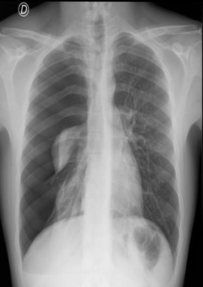
Figure 1: The final diagnosis maded after a clinical examination and chest X-ray.
The chest drainage was carried out, because of pneumothorax. About 30 minutes after pleural drainages tube placement, the patient started with thoracic pain and severe cough and was kept with Hypoxemia. We take immediately a control chest X-ray that revealed an alveolar infiltrate of the entire right lung field, which was interpreted as re-expansion pulmonary edema (REPE) (Figure 2).
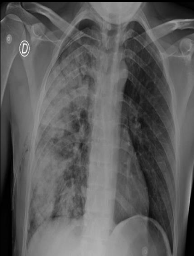
Figure 2: The chest drainage was carried out, because of pneumothorax.
We were treated successfully with supplemental oxygen therapy and methylprednisolone for 5 days, after the treatment the patient became asymptomatic and presented the following Chest X-ray (Figure 3).
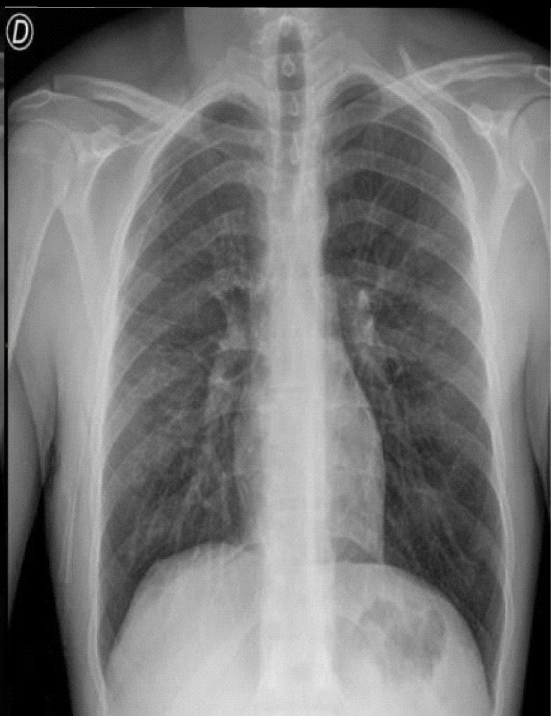
Figure 3: After the treatment the patient bécame asymptomatic and presented the following Chest X-ray.
Re-Expansion Pulmonary Edema (REPE) or unilateral shock lung is an infrequent clinical entity, with a low incidence rate. It can cause worsening in patients with a tension pneumothorax after placement of the pleural tube drainage.
The suspected diagnosis is made with an anamnesis and clinical history and requires confirmation by performing a chest X-ray. The treatment consists of bolus administration of methylprednisolone and supplemental oxygen, with complete resolution of symptomatology the in the majority of patients.
- Barril Merino C, Solovera R ME, Bannura Y F, Salas V P. Edema pulmonar agudo grave secundario a tratamiento de neumotórax espontáneo primario. Caso clínico [Pulmonary expansion edema during the management of a spontaneous pneumothorax. Report of one case]. Rev Med Chil. 2018 Nov;146(11):1343-1346. Spanish. doi: 10.4067/S0034-98872018001101343. PMID: 30725049.
- Schaer H, Roth F. Lungenoedem ex vacuo [Pulmonary oedema ex vacuo (author's transl)]. Anaesthesist. 1977 Oct;26(10):581-5. German. PMID: 579286.
- Mutz N, Benzer H, Coraim F, Pauser G, Schmid L. Lungenödem nach einseitigem Pneumothorax (Einseitige Schocklunge) [Pulmonary edema due to unilateral pneumothorax (unilateral shock lung) (author's transl)]. Prakt Anaesth. 1977 Oct;12(5):424-8. German. PMID: 917982.